Solitary Plasmocytoma of frontal bone presenting as an asymptomatic forehead lump: A case report
Published Web Location
https://doi.org/10.5070/D39s64p9t8Main Content
Solitary plasmocytoma of frontal bone presenting as an asymptomatic forehead lump
Jagajeevan Jagadeesan MRCS1, Deemesh Oudit MRCS1, Joseph Hardwicke MRCS1, Zakir Shariff MRCS1, Gavin McCoubrey MBChB1, Gareth Roberts FRCS2, Andrew Howcroft FRCS1
Dermatology Online Journal 12 (3): 24
Departments of Plastic and Reconstructive Surgery1 and Neurosurgery2, Royal Preston Hospital, Sharoe Green Lane, Fulwood, Preston. Lancashire, United Kingdom. drjjag@yahoo.comAbstract
Solitary plasmocytoma of bone is a rare type of plasma cell tumor. We present a case of a solitary extramedullary plasmacytoma of the frontal bone presenting as an asymptomatic forehead lump with clinically benign characteristics. This case highlights the need for a high index of suspicion when dealing with enlarging subcutaneous lumps of the forehead and scalp. The significance of this lies in the appropriate sequencing of investigations and the implementation of the necessary treatment regimen.
Lumps on the forehead are usually benign but sometimes may represent a metastatic or systemic malignancy presenting with the clinical features of a benign lesion. Solitary plasmacytoma of bone is a rare type of plasma cell tumor and is usually symptomatic. Here we present a case of solitary plasmacytoma of frontal bone that was entirely asymptomatic.
Clinical synopsis
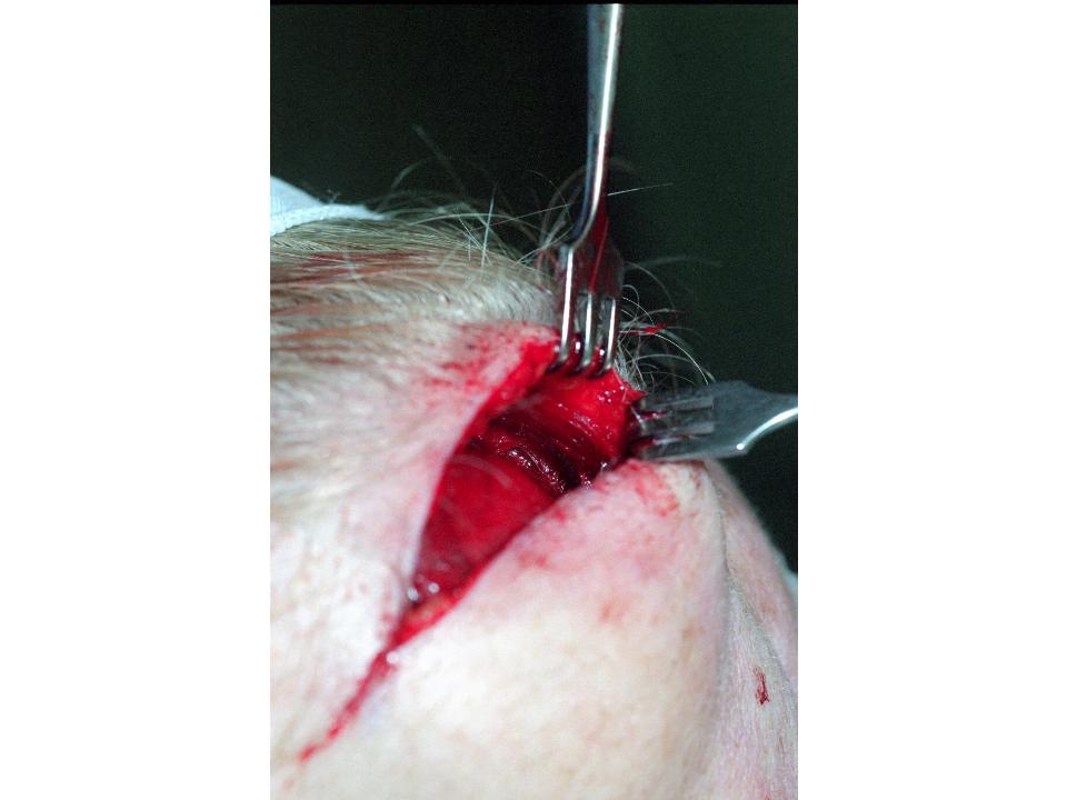 |
| Figure 1 |
|---|
| Intraoperatively, the lesion was found under cover of the frontalis muscle fibers. |
A 67-year-old female presented with an 18-month history of a painless, slowly enlarging lump on the right side of the forehead. The lesion was 5 cm in diameter, dome-shaped, nontender, rubbery in consistency and nonpulsatile. The skin overlying the lump was slightly mobile but it appeared to be tethered to the underlying structures. She reported no neurological symptoms and her only concern was the unsightly cosmetic appearance of the lesion. An initial diagnosis of a subfrontalis lipoma was made and arrangements were made for the lesion to be excised under local anasthetic. Intraoperatively she was found to have an ill-defined, soft, osteolytic lesion involving the frontal bone (Fig. 1). An FNA of the lesion was performed and the surgical wound was closed with a plan to proceed to further investigations and referral to the neurosurgeons. The FNA was inconclusive, raising the possibility of either a plasma cell tumor or a meningioma. A CT scan subsequently revealed evidence of a malignant tumor arising from the frontal bone, involving the inner table but with no evident intracranial extension. A secondary procedure was performed under the care of neurosurgeons and the tumor was excised along with the inner table of the underlying skull. Hispathological examination of the excised specimen confirmed the diagnosis of plasmacytoma of the frontal bone. The patient had an uneventful recovery from the procedure and was subsequently referred to a hematologist for further treatment.
Discussion
Plasmacytoma is a rare group of plasma cell neoplasm arising from the bone marrow or other soft tissue sites. Solitary bone plasmacytoma is an uncommon form of plasmacytoma and is localized to the involved bone [1]. They are usually symptomatic the most common symptom being pain as a result of bony erosion [2]. The other symptoms produced depend on the site of the lesion. Solitary plasmacytoma of skull may even present with Cranial nerve palsy depending on the site involved and the size of the lesion [3]. Radiotherapy has been considered the treatment of choice but most patients progress to multiple myeloma over a period of 3-10 years [4]. When combined with surgical excision either pre or post radiotherapy the curative rates are higher [5].
Forehead lumps either symptomatic or asymptomatic are a common presentation of the surgical outpatient. A significant proportion of subcutaneous lumps of the scalp and forehead are manifestations of malignancies (primary or secondary). Despite this, the index of suspicion that a scalp or forehead mass is malignant is usually low. This case emphasizes the need for high index of suspicion in cases of subcutaneous forehead nodules.
References
1. Di Micco P, Di Micco B. Up-date on solitary plasmacytoma and its main differences with multiple myeloma. Exp Oncol. Mar 2005; 27(1):7-122. Kyle RA: Monoclonal gammopathy of undetermined significance and solitary plasmacytoma. Implications for progression to overt multiple myeloma. Hematol Oncol Clin North Am. Feb 1997; 11(1): 71-87.
3. Z.Ustuner, M.Basaran, T. Kiris, et al. Skull Base Plasmacytoma in a Patient with Light Chain Myeloma. Skull Base. Aug 2003; 13(3): 167-171.
4. Aviles A, Huerta-Guzman J, Delgado S, et al. Improved outcome in solitary bone plasmocytoma with combined therapy. Hematol Oncol. Sept 1996: 14(3): 111 - 117.
5. P. Strojan, E. oba, J. Lamovec, A Munda. Extramedullary plasmacytoma: clinical and histopathologic study.International Journal of Radiation Oncology Biology Physics, July 2002; 53(3) : 692-701 .
© 2006 Dermatology Online Journal