Tripe palms associated with malignant acanthosis nigricans in a patient with gastric adenocarcinoma: A case report and review of the literature
Published Web Location
https://doi.org/10.5070/D37bh1h290Main Content
Tripe palms associated with malignant acanthosis nigricans in a patient with gastric adenocarcinoma: A case report and review
of the literature
Caterina Fabroni MD, Antonia Gimma MD, Carla Cardinali MD, Giovanni Lo Scocco MD
Dermatology Online Journal 18 (11): 15
Ospedale Misericordia e Dolce, Prato, ItalyAbstract
Tripe palms (TP) is a rare dermatologic condition. TP alone, or associated with malignant acanthosis nigricans (MAN), in most cases is a cutaneous paraneoplastic disorder and its recognition should prompt a full diagnostic work-up for an underlying malignancy. We report a case of a patient in whom the correct identification of TP and MAN has allowed early diagnosis of gastric cancer. Paraneoplasias are frequently the first sign of an underlying malignant tumor. Although relatively rare, they need to be recognized by dermatologists to make an early diagnosis and improve the prognosis related to the neoplasia.
Introduction
Tripe palms (TP) describes a rare dermatologic condition in which the skin of the palm becomes thick and velvety-white with pronounced folds in the lines of the hands [1]. The skin resembles boiled tripe. It is a sign of cancer in 95 percent of cases and its recognition should prompt a full diagnostic work-up for an underlying malignancy. TP is considered a cutaneous paraneoplastic syndrome that occurs predominantly in patients with solid tumors. Acanthosis nigricans (AN) is a brown to black, poorly defined, velvety hyperpigmentation of the skin folds. This dermatological condition, frequently related to insulin resistance, can also occur as a paraneoplastic syndrome. The term “malignant” AN (MAN) is used to suggest the association with malignancies [1, 2]. We describe a case of TP in conjunction with MAN that preceded the diagnosis of gastric carcinoma.
Case report
An 82-year-old woman was referred to our Dermatology Unit in Prato Hospital for a 5-month history of progressive hyperpigmentation and hyperkeratosis of her skin. She also complained of recent loss of weight, diarrhea, and weakness. There was no history of hematemesis, melena, hemoptysis, cough, or dyspnea. Her personal history was remarkable for mellitus diabetes type II.
 | 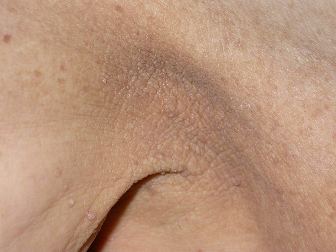 |
| Figure 1 | Figure 2 |
|---|
General examination revealed pallor. Dermatological examination showed diffuse hyperpigmentation with thickening of the skin of the face, neck, flexures, external genitalia, and anus (Figure 1). The hands showed enhanced ridges and velvety brownish-yellow hyperkeratosis involving the palmar surface of both hands (Figure 2). The soles were not affected. The tongue was fissured. Skin lesions were associated with moderate itching. Regional lymph nodes were not involved.
On admission, laboratory findings revealed a sideropenic anemia, increased values of CA 19-9 (90 U/ml; cut of 37 U/ml ), C-reactive protein (CRP:3.6 mg/dL), and erythrocyte sedimentation rate (ESR: 90 mm/h).
A skin biopsy, performed on the axillary surface, revealed hyperkeratosis, acanthosis, and dermal papillomatosis, consistent with the clinical diagnosis of acanthosis nigricans. With these clinical and histological findings, a diagnosis of MAN with TP was made and an extensive search for an underlying malignancy was initiated. Chest X-ray, mammogram, ultrasound abdomen, rectoscopy, and colonoscopy showed no evidence of malignancy. An upper gastrointestinal endoscopy and biopsy revealed gastric adenocarcinoma. Additional imaging excluded metastatic disease. Total gastrectomy was performed and two months after surgery the patient showed a marked clinical improvement of MAN and total disappearance of TP. After 6 months there was no sign of metastasis.
Discussion
The term “tripe palms” (TP) was introduced in the literature in 1977 by Jacqueline Clarke [2] and later popularized by Breathnach and Wells in 1979 [3]. TP is characterized by an acquired soft thickening, which assumes a mossy or velvety aspect associated with hypertrophy of the dermatoglyphics, making the palmar area pitted, furrowed, and rugose. In the most severe cases the palms have a honeycomb-like or cobbled appearance. This condition has also been referred to as acanthosis palmaris, pachydermatoglyphya, acquired palmoplantar keratoderma, or palmar acquired keratoderma [3-7]. Cohen et al. found TP to be associated with internal malignancy in more than 90 percent of 77 cases and is concomitant with MAN in 77 percent [4]. The internal malignancies associated include: gastrointestinal tract (30%), lung (20%), bladder (5%), breast (4%), cervical (4%), ovarian (4%), and renal (4%) [1-8]. Occasional cases have been reported of neoplasms originating in the colon, gallbladder, uterus, pancreas, prostate, and tongue; sarcomas, melanoma, and non-Hodgkin lymphoma have also been reported in patients with TP [9].
The onset of TP precedes malignancy in more than 40 percent, follows malignancy in 19 percent, or can be concurrent within one month of the diagnosis of malignancy in 37 percent of patients [1, 5]. The clinical manifestations of TP occur within a median of two months before the diagnosis of a malignant neoplasm [1, 2, 3].
TPs are frequently seen in conjunction with MAN [1, 2, 9]. In these cases, the underlying malignancy is most commonly stomach (35%) or lung (11%) cancer. Sometimes other cutaneous signs, such as the presence of multiple eruptive seborrheic keratoses (sign of Leser-Trelat), can be found in association with TP and MAN and can suggest an underlying malignancy [9].
Whether TP represents a distinct entity or merely the palmar manifestation of MAN remains unresolved. Some authors suggest that TP may be regarded as a separate entity when it occurs alone. However, the epidemiological, morphological, and histological characteristics of MAN and TP are similar [1, 2, 3, 10].
The cause of TP is not clearly understood but is thought to be related to the secretion of growth factors by the tumor cells that stimulate skin cells to proliferate. Transforming growth factor-α (TGF-α), structurally related to epidermal growth factor (EGF), has been considered possibly involved products involved. TGF-α could be a growth stimulating factor as suggested by some authors, a factor that increases the mitotic rate and consequently causes epithelial hyperplasia, which is responsible for the simultaneous appearance of several dermatological manifestation having as a common feature the hyperplasia of epithelial cells [5]. EGF receptors (EGFR) are found in keratinocytes, especially in the basal layer of the epidermis, and the number of receptors is increased in hyperproliferative skin disorders. Autocrine secretion of an EGF-like substance from a human skin squamous cell tumor line and from bronchial carcinoma has been demonstrated [11]. A similar pathogenic mechanism has been suggested by Haase for MAN [12]. Some authors suggested also that constitutive activation of Fibroblast growth factor receptor 3 (FGFR3) might have some relevance to the formation of MAN. Activation of EGFR, FGFR3, and insulin-like growth factor receptor could cooperate in the development of MAN [13, 14].
The diagnosis of TP and MAN is made from the characteristic clinical appearance whereas histological findings (characterized by an undulant epidermis with hyperkeratosis, acanthosis and papillomatosis) are not specific [1, 2, 3].
The clinical differential diagnosis of TP includes pachydermoperiostosis, hypertrophic pulmonary osteoarthropathy, acromegaly, thyroid acropachy, acrokeratosis paraneoplastica (Bazex syndrome), and keratosis palmaris et plantaris [1, 3].
There is no specific treatment for TP. Approximately 30 percent of cases resolve once the underlying cancer is treated. However, TP may persist for many years despite remission of the underlyng cancer [13].
We present this case to emphasize that cutaneous paraneoplasia (as TP or MAN) may be the first sign of an underlying cancer. Although relatively rare, these findings need to be recognized by dermatologists to make an early diagnosis and improve the prognosis related to the neoplasia.
References
1. Abreu Velez AM, Howard MS. Diagnosis and treatment of cutaneous paraneoplastic disorders. Dermatol Ther. 2010 Nov-Dec;23(6):662-75. [PubMed]2. Clarke J. Malignant acanthosis nigricans. Clin Exp Dermatol 1977; 2:167-170. [PubMed]
3. Breathnach SM, Wells GC. Acanthosis palmaris: tripe palms. A distinctive pattern of palmar keratoderma frequently associated with internal malignancy. Clin Exp Dermatol 1980; 5: 181-189. [PubMed]
4. Cohen PR, Grossman ME, Almeida L, et al. Tripe palms and malignancy. J Clin Oncol 1989; 7: 669-678. [PubMed]
5. Koulaouzidis A, Leiper K. Tripe palms or acanthosis palmaris. Intern Med J 2007; 37: 502. [PubMed]
6. Thappá DM, Garg BR, Venkateswaran S et al. Acanthosis palmaris: a marker of bronchogenic carcinoma. Acta Derm Venereol 1995; 75: 246. [PubMed]
7. Saeed H, Massarweh S. Images in clinical medicine. Hypertrophic pulmonary osteoarthropathy and tripe palms. N Engl J Med. 2012 Jan 26;366(4):360. [PubMed]
8. Patel S, Zirwas M. Acquired palmoplantar keratoderma. Am J Clin Dermatol 2007; 8: 1-11. [PubMed]
9. Pentenero M, Carrozzo M, Pagano M, Gandolfo S. Oral acanthosis nigricans, tripe palms and sign of Leser-Trélat in a patient with gastric adenocarcinoma. Int J Dermatol 2004;43:530-2. [PubMed]
10. Costa MC, Martinez NS, Belicha MG, et al. Florid cutaneous papillomatosis and acanthosis nigricans maligna revealing gastric adenocarcinoma. An Bras Dermatol. 2011 May-Jun;86(3):573-7
11. Gorisek B, Krajnc I, Rems D, Kuhelj J. Malignant Acanthosis Nigricans and Tripe Palms in a patient with Endometrial Adenocarcinoma - a case report and review of literature. Gynecology Oncology 65,539-542 (1997) [PubMed]
12. Douglas F, McHenry PM, Dagg JH, MacBeth FM, Morley WN. Elevated levels of epidermal growth factor in a patient with tripe palms. Br J Dermatol. 1994 May;130(5):686-7. [PubMed]
13. Haase I, Hunzelmann N. Activation of Epidermal Growth Factor Receptor/ERK Signaling Correlates with Suppressed Differentiation in Malignant Acanthosis Nigricans. J Invest Dermatol. 2002 May;118(5):891-3. [PubMed]
14. Hida Y, Kubo Y, Nishio Y, Murakami S, Fukumoto D, Sayama K, et al. Malignant acantosis nigricans with enhanced expression of fibroblast growth factor receptor 3. Acta Derm Venereol. 2009;89:435-7. [PubMed]
15. Berardesca E, Del Forno C, Vignini M: Uselfulness of etretinate treatment in paraneoplastic palmoplantar hyperkeratosis. Br J Dermatol. 117: 132-133, 1987. [PubMed]
© 2012 Dermatology Online Journal