Cutaneous mastocytosis with systemic involvement mimicking clinical and dermatoscopically multiple melanocytic nevi
Published Web Location
https://doi.org/10.5070/D372x522rfMain Content
Letter: Cutaneous mastocytosis with systemic involvement mimicking clinical and dermatoscopically multiple melanocytic nevi
Enrique Gutiérrez-González MD, Manuel Ginarte PhD, Jaime Toribio PhD
Dermatology Online Journal 17 (11): 15
Department of Dermatology, Complejo Hospitalario Universitario, Faculty of Medicine, Santiago de Compostela, SpainAbstract
Mastocytosis can sometimes resemble other skin conditions, especially pigmented ones, not only clinically but also dermatoscopically. We report the case of a woman with the diagnosis of cutaneous mastocytosis mimicking multiple melanocytic nevi. Melanocytic stimulation can be induced by high levels of stem cell factor. The progressive increase in the number of pigmented lesions in a patient should lead us to perform a biopsy to search for mastocytosis.
Mastocytosis can sometimes resemble other skin conditions, especially pigmented ones. We report the case of a woman diagnosed with cutaneous mastocytosis with systemic involvement (bone marrow infiltration) whose lesions, on dermatoscopic examination, showed a fine, regular pigmented network. This finding can be explained by the increased number of melanocytes in the basal layer and also increased melanin production, related to stem cell factor (SCF) stimulation.
 |  |
| Figure 1 | Figure 2 |
|---|---|
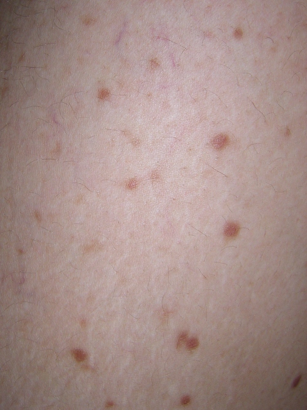
| Figure 1. Brown macules scattered over the thighs Figure 2. Dermatoscopy showing brown reticular lines (pigment network) | |
 |
| Figure 3 |
|---|
| Figure 3. Hyperpigmentation of keratinocytes in the basal layer and metachromasia of mast cells (Toluidin blue, x40) |
A 32-year-old woman presented in our clinic with a significant increment in the number of her “moles” over the last year, but otherwise the lesions were asymptomatic. She was in good health and was not taking any medication. On physical examination, multiple brown macules were present all over the trunk and extremities, but especially on the thighs (Figure 1). Darier sign was negative. Dermatoscopic examination of the lesions revealed brown reticular lines, drawing up a fine, regular pigment network (Figure 2). A biopsy of one of the lesions was performed, showing an almost normal epidermis, with a slight increase of melanocytes and melanin deposits on the basal layer (Figure 3). In the dermis there was a very dense cellular infiltrate, consisting of round cells with cytoplasm full of granules that stained metachromatically with toluidine blue and Giemsa stain (Figure 3). Diagnosis of cutaneous mastocytosis was then established. A full blood count and peripheral blood smear revealed no abnormality, although tryptase serum levels were elevated. Radiographies of several bones failed to show any lesions. However, a bone marrow biopsy was performed, showing bone marrow infiltration by mast cells, with an atypical inmunophenotype (CD2 and CD25+) as well as c-Kit mutation in 10-20 percent of the alleles.
Mastocytosis is a disorder characterized by mast cell proliferation and accumulation in several organs and tissues, the skin being the most common. The diagnosis of cutaneous mastocytosis is based on the finding of a dermal infiltrate composed of mast cells that can be recognized on skin sections, better visualized by the use of special stains such as Giemsa or Toluidine blue [1]. The c-Kit proto-oncogene codes for the transmembrane tyrosine kinase receptor of the SCF. An activating mutation of c-Kit leads to a ligand independent auto-phosphorylation. As a consequence, this produces autonomous mast cell proliferation [1]. The accumulation of and increase in the soluble SCF levels may lead to a higher melanocytic proliferation, melanin pigment production, and melanin deposit in basal keratinocytes because of the SCF receptors also present in melanocytes [2]. This may explain the presence of the clinical brownish color and dermatoscopic (pigment network) findings. Pigment network is a feature usually found in melanocytic lesions and considered as diagnostic, but non-exclusive because it has been described in other lesions like seborrheic keratoses or dermatofibroma [2, 3, 4]. Histologically it corresponds to the presence of increased amounts of melanin in the basal layer of the epidermis. Dermatoscopy, can be of great help in recognizing these changes in pigmentation. Few descriptions showing the dermatoscopic findings in cutaneous mastocytosis have been previously reported [2, 5].
In conclusion, cutaneous mastocytosis can resemble melanocytic nevi, not only clinically, but also dermatoscopically. A progressive increase in the number of pigmented lesions in a patient should lead us to perform a biopsy to search for mastocytosis. We assert that cutaneous mastocytosis should be considered in the differential diagnosis of lesions presenting with pigment network on dermatoscopy.
References
1. Hartmann K, Henz BM. Mastocytosis: recent advances in defining the disease. Br J Dermatol 2001; 144(4): 682-95. [PubMed]2. Akay BN, Kittler H, Sanl H, Harmankaya K, Anadolu R. Dermatoscopic findings of cutaneous mastocytosis. Dermatology 2009; 218(3): 226-30. [PubMed]
3. Arpaia N, Cassano N, Vena GA. Dermoscopic patterns of dermatofibroma. Dermatol Surg. 2005 Oct; 31(10): 1336-9. [PubMed]
4. De Giorgi V, Massi D, Stante M, Carli P. False “melanocytic” parameters shown by pigmented seborrheic keratoses: a finding which is not uncommon in dermoscopy. Dermatol Surg 2002 Aug; 28(8): 776-9. [PubMed]
5. Arpaia N, Cassano N, Vena GA. Lessons on dermoscopy: pigment network in non melanocytic lesions. Dermatol Surg 2004 Jun; 30(6): 929-30. [PubMed]
© 2011 Dermatology Online Journal