Ulcerating necrobiosis lipoidica resolving in response to cyclosporine-A
Published Web Location
https://doi.org/10.5070/D36f7981ksMain Content
Ulcerating necrobiosis lipoidica resolving in response to cyclosporine-A
Kevin Smith, M.D.
Dermatology Online Journal: 3(1): 2
AbstractNecrobiosis lipoidica often fails to respond adequately to therapy with topical and intralesional corticosteroids, or to systemic medications like niacinamide and pentoxifylline (Trental). On the basis of unpublished work which showed a predominance of T helper cells in lesions of necrobiosis lipoidica, and recalling the case of a woman whose necrobiosis lipoidica improved after she was started on cyclosporine for a renal transplant, systemic cyclosporine was successfully used in the cases of two young women who had insulin-dependent diabetes and were disfigured by severe, ulcerating necrobiosis lipoidica on the anterior lower legs. Response to treatment was monitored with photographs. In both cases the ulcers resolved, and remained in remission after cyclosporine was stopped. |
IntroductionNecrobiosis lipoidica (NL) is closely associated with diabetes. About 660f patients with NL have overt diabetes, and additional 4% have an abnormal glucose tolerance test, and 110f those with a normal glucose tolerance test have a family history of diabetes. NL occurs in about 0.30f patients with diabetes, with a female:male ratio of 3:1. NL can occur simultaneously with granuloma annulare (GA), which has a similar histology to NL and which is occasionally associated with diabetes. Many patients with NL may demonstrate diabetic end organ damage, including nephropathy, neuropathy and retinopathy.(1)NL often fails to respond adequately to conventional therapy with topical and intralesional corticosteroids, or to systemic medications like niacinamide and pentoxifylline (Trental). On the basis of unpublished work with Arnold Schroeter and Richard Winkelmann which showed a predominance of T-helper cells in lesions of necrobiosis lipoidica, and recalling the case of a woman whose necrobiosis lipoidica improved after she was started on cyclosporine for a renal transplant, I suggested a trial of treatment with cyclosporine to two young women who had insulin-dependent diabetes and were disfigured by severe, ulcerating necrobiosis lipoidica on the anterior lower legs. MethodsResponse to therapy was monitored by serial photographs of the involved areas. Both the patients and myself knew that active cyclosporine was being administered (there was no placebo control). Patients were given printed instructions on the correct use of cyclosporine. Patients were instructed to monitor blood glucose daily, and they reported that there were no changes from usual while taking cyclosporine. Creatinine and blood pressure were monitored once a week for the first month, then twice a month while the patients were taking cyclosporine.Case 1: Female with insulin dependent diabetes diagnosed at the age of 14 in 1987, and biopsy proven NL on the lower legs since 1990. The lesions on the lower legs did not respond adequately to trials of treatment with:
|
|
| FIGURE 1 | FIGURE 2 |
|---|
|
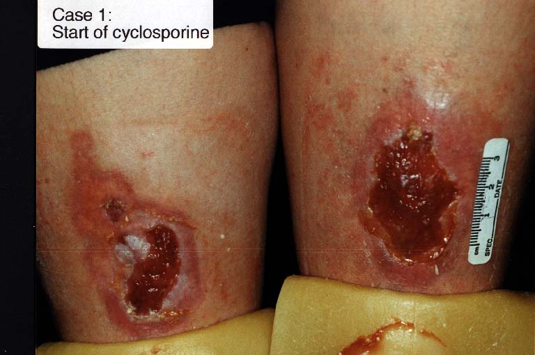
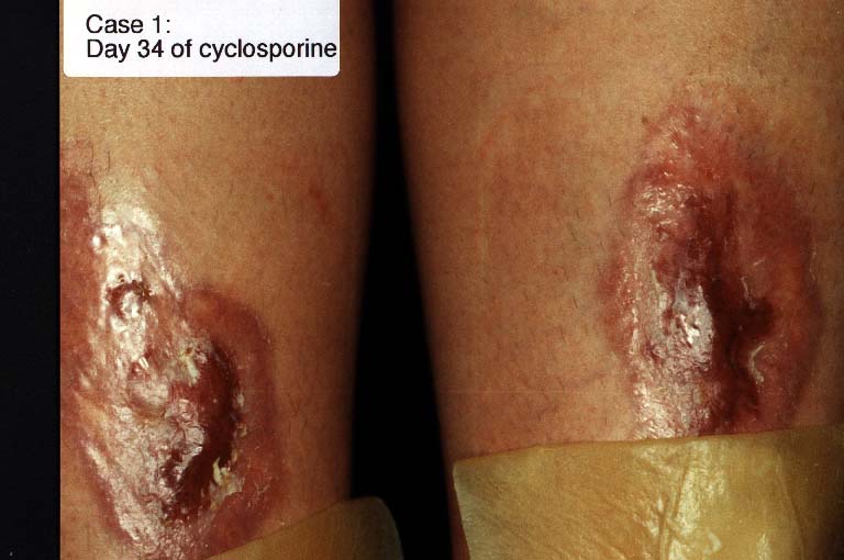
Figure 1 (left): Case 1: start of cyclosporine. Figure 2 (right): Case 1: day 34 of cyclosporine |
|
| FIGURE 3 | FIGURE 4 |
|---|
|
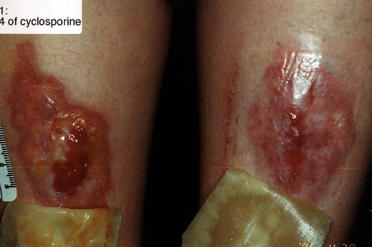
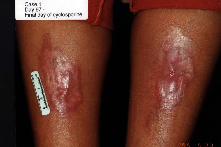
Figure 3 (left): Case 1: day 64 of cyclosporine Figure 4 (right): Case 1: day 97 - final day of cyclosporine |
|
| FIGURE 5 | FIGURE 6 |
|---|
|
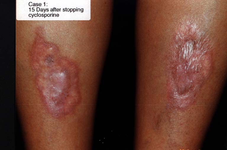
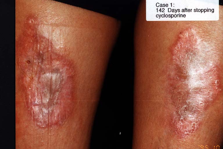
Figure 5 (left):Case 1: 15 days after stopping cyclosporine Figure 6 (right): Case 1: 142 days after stopping cyclosporine |
Case 2: Female with insulin-dependent diabetes diagnosed at the age of 13 in 1977, and NL on the lower legs since 1983. Ulceration in the lesions of NL started in 1989, and did not respond adequately to:
There was a transient elevation of the serum creatinin to 189 µmol/L in October 1995 and Sandimmune Neoral was stopped, with return of the serum creatinin to normal over a period of several months. Several 1 cm erosions developed in the lesions of NL in December 1995, perhaps because the patient had started to smoke in December. Smoking was stopped, and the NL has been kept in remission since December 1995 with PUVA twice a week and Trental 800 mg tid. DiscussionThe following lines of evidence led to this trial of treatment with cyclosporine-A in the cases presented here:
Because of the potential for nephrotoxicity, in particular in diabetics who may already have poor renal function secondary to diabetic nephropathy , close monitoring of serum creatinin is essential, in particular when the patient is taking medications such as erythromycin (Case Two) which may increase the blood level of cyclosporine. ConclusionCyclosporine-A is useful in the management of severe ulcerating NL, and long-term treatment with cyclosporine was not needed in these cases. Acknowledgement: Sandimmune Neoral was provided by Sandoz Canada. The author has acted as a speaker for Sandox (Novartis) Corporation. References1. Demis: Clinical Dermatology; Volume 1, Unit 4-8:1-7, revision CD-94.Lippincott-Raven, Philadelphia, 1997.2. Honig B, Morrison WL, Karp D: Photochemotherapy beyond psoriasis. J Am Acad Dermatol 31:775-790, 1994. 3. Handfield-Jones S, Jones S, Peachey R: High-dose nicotinamide in the treatment of necrobiosis lipoidica. Br J Dermatol 118:693-696, 1988. 4. Goette DK: Resolution of necrobiosis lipoidica with exclusive clobetasol propionate treatment. J Am Acad Dermatol 22:855-856, 1990. 5. Sparrow G, Abell E: Granuloma annulare and necrobiosis lipoidica treated by jet injector. Br J Dermatol 93:85-89, 1975. 6. Petzelbauer P, Wolff K Tappeiner G: Necrobiosis lipoidica: Treatment with systemic corticosteroids. Br J Dermatol 126 :542-545, 1992. |
© 1997 Dermatology Online Journal for use in electronic version, otherwise copyright retained by the author(s) |