Pseudoxantoma elasticum-like dermal elastolysis: A case report
Published Web Location
https://doi.org/10.5070/D35pd5f1r9Main Content
Pseudoxantoma elasticum-like dermal elastolysis: A case report
Verónica López MD1, Angeles Revert MD1, Nuria Santonja MD2, Esperanza Jordá MD PhD1
Dermatology Online Journal 17 (4): 13
1. Department of Dermatology2. Department of Pathology
Hospital Clínico Universitario de Valencia, Valencia, Spain
Abstract
Elastic fibers are components of dermal connective tissue that can be affected in several acquired disorders. Recently, a new entity known as pseudoxanthoma-like papillary dermal elastolysis has been described. We present a case in a 61-year-old woman.
Elastic fibers are components of dermal connective tissue that can be affected in several acquired disorders. Recently, a new entity known as pseudoxanthoma-like papillary dermal elastolysis has been described [1]. It is a rare condition described in less than twenty patients and presents as multiple, tiny, flesh-color or yellowish papules with a cobblestone-like distribution over the posterior and lateral sides of the neck, axillae, flexor forearms, lower abdomen, and inframmary folds of women between 60 and 80 years old [1, 2].
 |  |
| Figure 1 | Figure 2 |
|---|---|
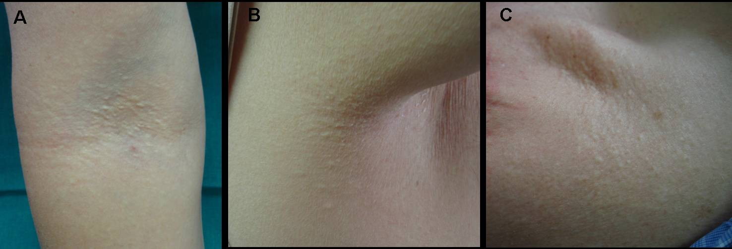
| Figure 1. Multiple flesh-colored, 2mm cobblestone-like papules over the antecubital fossae (A), axillae (B), and the neck
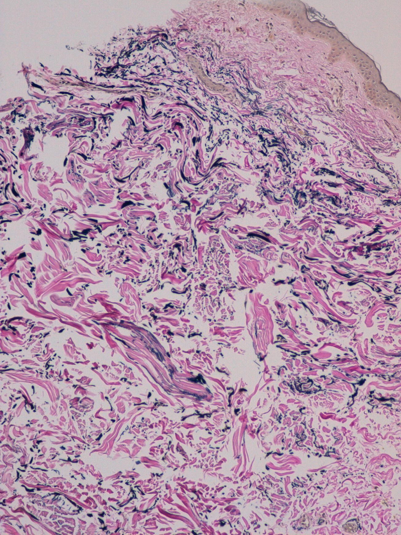
(C). Figure 2. Low power view showing elastic fibers in the papillary dermis with abnormal distribution in the reticular dermis (Orcein, x10) | |
A 61-year-old woman was referred to our department with a history of multiple asymptomatic lesions over the lateral and posterior side of her neck, axillae, and antecubital fossae of several years of evolution. She did not note similar lesions in any member of her family. Physical examination demonstrated multiple flesh-colored, 2 mm non-follicular papules grouped to form cobblestone-like plaques over her neck, axillae, and antecubital fossae (Figure 1). Mucosal involvement was not present. She did not have any cardiovascular, gastrointestinal, or ocular symptoms. One of the lesions was biopsied.
The histological examination with hematoxilyn-eosin stain showed no abnormalities. The orcein stain demonstrated loss of elastic tissue in the papillary dermis with an anomalous distribution and morphology of elastic fibers in the reticular dermis (Figure 2).
A complete blood test, an electrocardiogram, echocardiography, and an ophthalmological examination were performed with normal results. A diagnosis of pseudoxantoma elasticum-like dermal elastolysis was made.
Pseudoxantoma elasticum-like dermal elastolysis (PELDE) was first described in 1992 by Rongioletti et al. Since then only 19 cases have been reported [1, 2]. It is a rare, acquired condition that tends to affect women between 60 and 80 years old [3]. Clinically, it is characterized by multiple, tiny, flesh-color or yellowish, non-follicular papules coalescing to form cobblestone-like plaques. It usually presents with a symmetrical distribution over the posterior and lateral sides of the neck, axillae, flexor forearms, lower abdomen, and inframmary folds [4]. Unlike, pseudoxantoma elasticum, PELDE is not associated with any systemic manifestations. Histopathological findings consist of an atrophic epidermis and band-like loss of elastic tissue in the papillary dermis [5]. Clumping and fragmentation of elastic fibers, loss of elaunin and oxylatan fibers in the papillary dermis of lesional skin, and granular and fibrillar degeneration of elastic fibers in perilesional areas may also be seen [4].
The differential diagnosis includes pseudoxantoma elasticum (PXE), white fibrous papulosis of the neck, upper dermal and middermal elastolysis, late-onset focal dermal elastosis, and perforating periumbilical calcific elastosis. PXE is a rare inherited disorder characterized by fragmentation and calcification of elastic fibers with association of systemic symptoms, including ocular and cardiovascular manifestations. White fibrous papulosis of the neck presents clinically as white papules on the neck, with histological changes consisting of areas of thickened collagen bundles in the papillary and midreticular dermis. Upper dermal elastolysis is characterized by a papular eruption on the neck, shoulders, upper part of the chest, and back with selective loss of elastic tissue in the upper dermis. Mid-dermal elastolysis manifests clinically as patches and plaques of finely wrinkled skin with focal loss of elastic tissue in the midreticular dermis upon histopathological examination. Late-onset focal dermal elastolysis occurs in older patients as multiple yellowish papules that may coalesce over the neck, axillae, groin, and antecubital and popliteal fossae. Histologically, increased normal-appearing elastic fibers in the mid and deep reticular dermis are observed. Perforating periumbilical calcific elastosis appears as coalescing keratotic papules with transepidermal elimination of calcified, distorted elastic fibers.
To date, there is no effective treatment for pseudoxantoma elasticum-like dermal elastolysis.
In conclusion, there are few cases of pseudoxantoma elasticum-like dermal elastolysis reported in the literature. We have described a new case of PELDE in a 61-year-old woman.
References
1. Rongioletti F, Rebora A. Pseudoxantoma elasticum-like papillary dermal elastolysis. J Am Acad Dermatol. 1992; 26:648-50. [PubMed]2. Monteagudo B, Cabanillas M, Used-Aznar M. Elastólisis dérmica papilar similar a seudoxantoma elástico: presentación de un caso y revisión de la literatura. Piel. 2009; 24:17-9.
3. El-Charif MA, Mousawi AM, Rubeiz NG, Kibbi AG. Pseudoxanthoma elasticum like papillary dermal elastolysis: a report of two cases. J Cutan Pathol 1994; 21:252-5. [PubMed]
4. Byun JY, Do MO, Kim SH, Choi HY, Myung KB, Choi YW. Pseudoxanthoma elasticum-like papillary dermal elastolysis developed in early middle age. J Dermatol. 2007; 34:709-11. [PubMed]
5. Vargas-Diez E, Penas PF, Fraga J, Aragues M, Garcia-Diez A. Pseudoxanthoma elasticum-like papillary dermal elastolysis. A report of two cases and review of the literature. Acta Derm Venereol 1997; 77:43-5. [PubMed]
© 2011 Dermatology Online Journal