Furuncular myiasis: Unusual case of African
Published Web Location
https://doi.org/10.5070/D39xb18658Main Content
Furuncular myiasis: Unusual case of African Dermatobia hominis
R Frikh MD1, N Hjira MD1, M Frikh MD2, N Baba MD1, M Ghfir MD1, B Lmimouni MD2, O Sedrati MD1
Dermatology Online Journal 15 (9): 11
1. Department of Dermatology2. Department of Parasitology
Military Hospital Mohamed V, Rabat, Morocco. rfrikh@gmail.com
Abstract
Human cutaneous myiasis is a common disease in endemic tropical zones. Increased international travel has produced increases in imported cases. We present an unusual patient with myiasis infestation of the leg caused by Dermatobia hominis, which manifested after returning from the Democratic Republic of Congo. This particular infestation has not been reported in Morocco prior to this case. Furuncular cutaneous miyasis must be considered when travellers exhibit draining nodules. Medical treatment consists of occlusion of the furuncular punctum with vaseline to stimulate extrusion of the larva or surgical debridement under local anesthesia.
Introduction
The infestation of tissues and organs by larvae of certain Dipteran flies is termed, myiasis. Myiasis is a worldwide phenomenon that is related to the latitude and the lifecycle of certain species of flies. Cases of myiasis are found most commonly in tropical and subtropical zones of Africa and the Americas [1]. The flies responsible for the condition prefer a warm and humid environment. Therefore, myiasis is limited to the summer months in temperate zones, but may occur all year in the tropics.
In the last two decades there has been an increase in travel to exotic destinations. Several reports detail patients suffering from infestations by Dermatobia hominis after returning from tropical parts of South America [1, 2, 3, 4] and by Cordilobia anthropophaga among those returning from Africa [5].
We present an unusual infestation of the leg caused by Dermatobia hominis that occurred after our patient returned from the Democratic Republic of Congo.
Case presentation
A 47-year-old man presented with skin lesions on his left leg five days after returning from a military mission in the Democratic Republic of Congo. The patient noted erythema, itching, and subsequent burning and tenderness in one nodule on the leg that had begun to drain. He reported that there were numerous insects and he affirmed to having been bitten by insects in the field. The patient had no constitutional symptoms.
 |  |
| Figure 1 | Figure 2 |
|---|---|
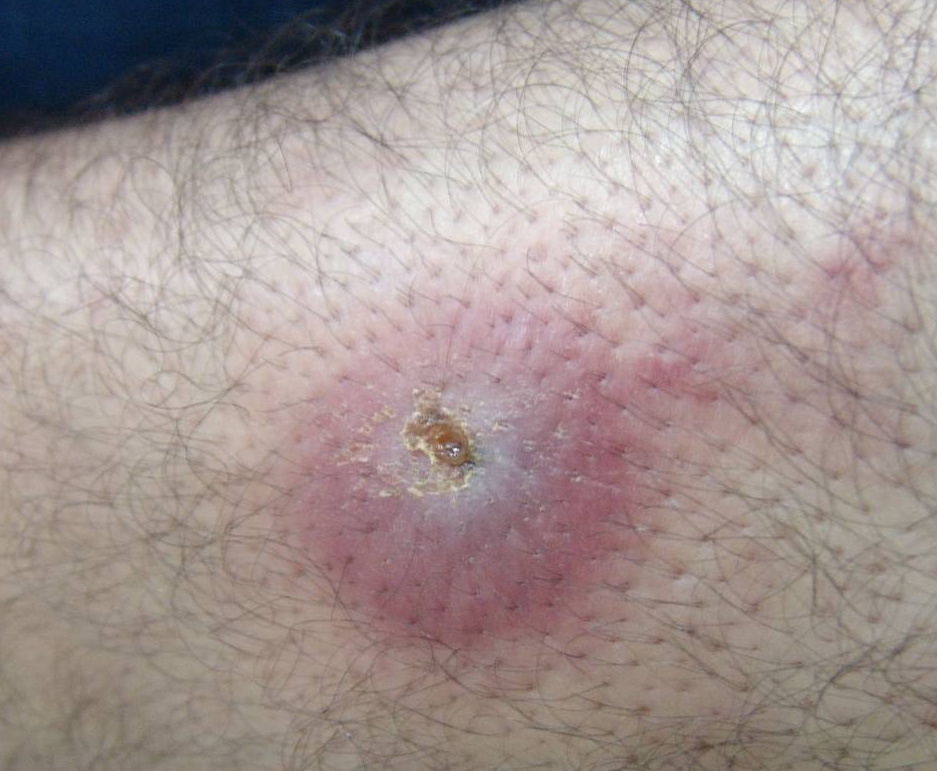
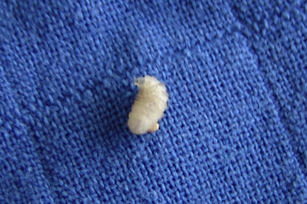
| Figure 1. Representative photograph of furonculoid lesion of the leg, with central pore, which can be confused with a simple
carbuncle. Figure 2. Larvae of myiasis after extraction. | |
Physical examination revealed one nodule on the middle third of the left leg, measuring 3 cm in diameter, with surrounding erythema and a central pore; mild local lymphangitis was apparent (Fig. 1). The results of routine blood studies were normal.
The pores were occluded with vaseline and the nodule became more swollen. After a few hours, two larvae were delivered (Fig. 2); the nodule resolved with additional oral treatment for secondary bacterial infection (flucloxacilline 3 g per day).
 |  |
| Figure 3 | Figure 4 |
|---|---|
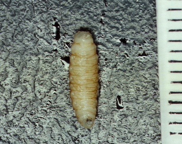
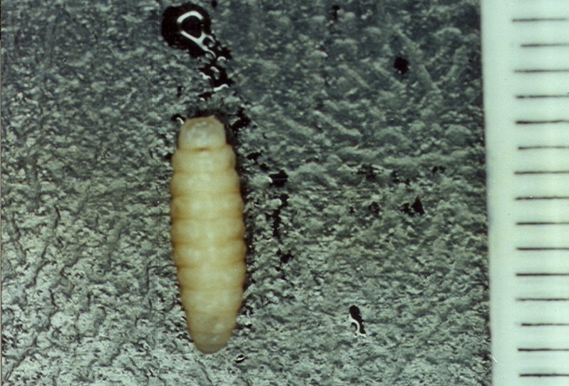
| Figure 3. Photomicrograph of third stage larva of Dermatobia hominis (ventral face), removed from the lesion described. Figure 4. Photomicrograph of dorsal face of larvae | |
The larvae were identified as Dermatobia hominis by a parasitologist (Figs. 3 & 4). The identification was made by comparing both anterior and posterior peritremes (spiracles openings) as well as body spinulation of the two larvae. The rows of dark brown, caudally pointing, barb-like spines indicated the larva was Dermatobia hominis.
Discussion
Myiasis is the term derived from the Greek word for fly, or myia. Myiasis describes infestations of warm-blooded hosts by flies, which usually involve the larval or pupal stages of the flies. In some instances, myiasis is beneficial to the host; larvae of some species clean and digest necrotic tissue. At one time, this was an accepted therapeutic regimen for dirty wounds [1].
The most impressive forms of myiasis relate to the appearance of furuncular varieties in the skin, which are being seen with increasing prevalence. The rapid pace of present-day air travel allows for the development of symptoms shortly after the tourist's return home.
Most commonly, skin involvement with myiasis, occurs as a result of infestation by Dermatobia hominis in patients returning from South America [2, 3, 4], and Cordilobia anthropophaga among those returning from Africa [5]. To the best of our knowledge, no case of furuncular myiasis due to D. hominis has yet been reported in a patient returning from Africa.
The life cycle of D. hominis (the human botfly) begins when the ova from the fly are laid directly onto the host, either in viable or necrotic tissue. The female fly can also lays eggs on the cephalothorax of certain mosquitoes. The mosquitoes then bite the human host; at this time, the eggs hatch and the larvae drop off and penetrate the human host [1, 6, 7].
Myiasis may have multiple presentations related to the different life cycles of the various Diptera genera. Furuncular cutaneous miyasis, which is caused by the human botfly, causes furunculoid (boil-like) lesions. After hatching, the larvae enter the skin and remain stationary. A pruritic papule occurs within 24 hours after contact, which enlarges to nodule of 1 to 3 cm in diameter. These lesions can be painful and may become crusted and purulent, as in our patient. There is typically an opening at the top of these boil-like swellings for entry of oxygen. Via this opening, the larva obtains oxygen. Dermatobia hominis has characteristic black, backward-pointing spines that anchor it in the host's skin and make its removal difficult [6, 7].
After 2 to 3 months, the larvae leave the skin and drop to the ground to pupate. The most commonly reported myiasis-causing species is Dermatobia hominis; the mosquito undoubtedly increases the spread of D. hominis larvae [8, 9].
The most likely disease in the differential diagnosis is bacterial infection. Other possible conditions include include leishmaniasis or very early dracunculosis (Guinea worm) [1, 7, 10].
Appropriate evaluation and correct diagnosis requires an awareness of the clinical presentation and careful examination. Occasionally laboratory tests may be helpful, including evaluation of blood/tissue eosinophilia, mainly in chronic cases of cutaneous myiasis. Biopsy with special stains to look for organisms may also be a useful diagnostic step. In terms of diagnostic testing, ultrasound can assist in the diagnosis, determine the size of larva, and evaluate the response to treatment [7, 11]. The morphology of the spiracles can allow identification of the larvae, with comparison of anterior and posterior peritremes. Cordylobia anthropophga has characteristic posterior spiracles. However, Dermatobia hominis has rows of dark brown, and caudally pointing, barb-like spines.
The goal of treatment is to remove the larva and treat any associated bacterial infection with antibiotics. The traditional treatment in areas where myiasis is endemic (Mexico, South and Central America) is occlusion of the furuncular punctum with pork fat to stimulate extrusion of the larva by suffocation, forcing it to wriggle out. Substances used include oil, petroleum jelly, butter, and liquid paraffin [1, 4]. The larvae in furuncular cutaneous myiasis should not be forcibly removed through its punctum because its tapered shape with rows of spines and hooks prevents simple extrusion [8]. Surgical debridement under local anesthesia is curative, although a foreign body response can occur if parts of the larvae remain. Given that myiasis can be a portal of entry for Clostridium tetani, vaccination should be considered with this infestation. An alternative treatment for all types of myiasis would be oral ivermectin. [12].
Conclusion
Myiasis is a self-limiting infestation, with the vast majority of cases having minimal morbidity. Indeed, removal of the larvae allows more rapid healing and clinches the diagnosis. In addition, the patient will feel much better to know that the maggot has been removed from his or her body. In endemic countries, this is a familiar condition that is easily recognized. But for the traveller, an emergency room physician at home may be unfamiliar and may need to seek expert consultation.
References
1. Millikan LE. Myiasis. Clin Dermatol 1999;17:191-5. [PubMed]2. Desruelles F, Delaunay P, Marty P, Del Giudice P, Mantoux F, Le Fi choux Y, Ortonne J.P. Myiasis caused by Dermatobia hominis after an organized tour to Amazonia. Presse Med 1999;28:2223- 5. [PubMed]
3. Maier H, Honigsmann H. Furuncular myiasis caused by Dermatobia hominis, the human botfly. J Am Acad Dermatol 2004;50(Suppl 1): S26- S30. [PubMed]
4. Marty FM, Whiteside KR. Myiasis due to Dermatobia hominis (Human Botfly). N Engl J Med 2005;352: e21. [PubMed]
5. Adisa CA, Mbanaso A. Furuncular myiasis of the breast caused by the larvae of the Tumbu fly (Cordylobia anthropophaga). BMC Surg. 2004;4:5. [PubMed]
6. Lemon MA, Aeling JL. Furuncular Myiasis. N Engl J Med 2000; 342:937. [PubMed]
7. Cestari TF, Pessato S, Ramos-e-Silva M. Tungiasis and myiasis. Clin Dermatol 2007; 25:158-64. [PubMed]
8. Meinking TL, Burkhart CN, Burkhart CG. Changing paradigms in parasitic infections: common dermatological helminthic infections and cutaneous myiasis. Clin Dermatol 2003;21:407-16. [PubMed]
9. Ruiz-Martinez I, Gomez F, Perez JM. The role of botfly myiasis due to Dermatobia homini L.Jr. (Diptera: Cuterebridae) as a predisposing factor to New World screwworm myiasis (Cochliomyia hominivorax coquerel) (Diptera:Calliphoridae). Ann NY Acad Sci 1996;791: 434 – 42. [PubMed]
10. Johnston M, Dickinson G. An unexpected surprise in a common boil. J Emerg Med 1996;14:779–81. [PubMed]
11. Quintanilla-Cedillo MR, Leon-Urena H, Contreras-Ruiz J, Arenas R. The value of Doppler ultrasound in diagnosis in 25 cases of furunculoid myiasis. Int J Dermatol. 2005;44(1):34-7. [PubMed]
12. Burkhart CN. Ivermectin: an assessment of its pharmacology, microbiology, and safety. Hum Vet Med 1999; s42:30–35. [PubMed]
© 2009 Dermatology Online Journal