Retronychia: A rare cause of chronic paronychia
Published Web Location
https://doi.org/10.5070/D39qr4059jMain Content
Retronychia: A rare cause of chronic paronychia
Ines Zaraa1,2, Rym Kort1, Mourad Mokni1,2, Amel Ben Osman1,2
Dermatology Online Journal 18 (6): 9
1. Dermatology, La Rabta Hospital, Tunis, Tunisia2. Faculté de médicine de Tunis, Université El Manar, Tunis, Tunisia
Abstract
Retronychia, described in 1999, is a rare entity of ingrown toenails. Embedding of the nail into the proximal nail fold (PNF) leads to chronic inflammatory changes. Herein, we report a new case that exhibited persistent paronychia in a 23-year-old woman. Retronychia usually does not recur once treated with avulsion. It should be suspected in the event of chronic proximal paronychia.
Case synopsis
A 23-year-old woman with no pertinent medical history presented with a 4-month period of painful swelling of her right great toenail. There was no history of a traumatic precipitating event except the wearing of tight shoes. She did not respond to topical steroids and systemic antibiotics.
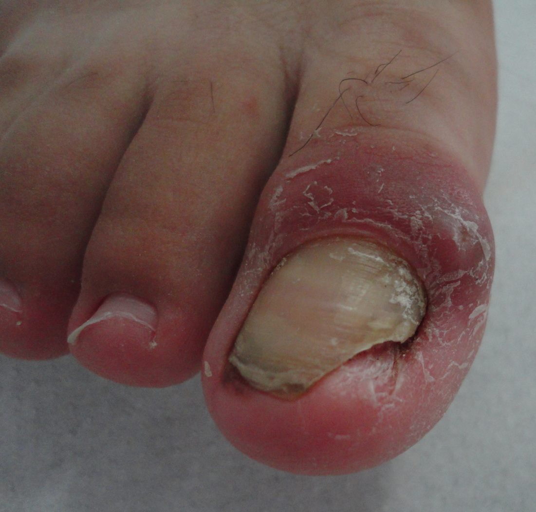
On examination, the right great toenail exhibited yellowish discoloration with distal onycholysis. The PNF was swollen with marked inflammation (Figure 1). The diagnosis of retronychia was made based on medical history and clinical findings. An avulsion of the nail under digital block anesthesia was performed. The nail was freed from the PND and lifted out.
 |  |
| Figure 1 | Figure 2 |
|---|---|
| Figure 1. Extensive paronychia at the time of presentation: retronychia starts with disruption of the longitudinal growth
of the nail because of an insult, and then complete with the growth of a new nail, the old one is pushed upwards and backwards.
This leads to embedding of the top nail into the ventral aspect of the proximal nail fold and subsequent inflammation of the
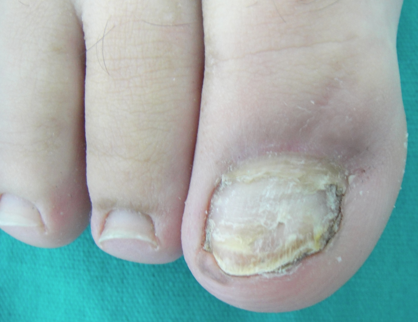
periungual skin Figure 2. Healthy looking nail, 9 months after surgical avulsion of the top nail. | |
The patient was without complaint within 10 days after surgery. Nine months later, the nail had regrown normally (Figure 2).
Retronychia is a latino-greek term which signifies backwards for retro and nail for onychia [3]. Since its first description, fewer than 40 cases have been cited in the literature [1, 2, 3]. It usually affects young adults (mean age about 39 years) with a female preponderance [3]. It starts with the disruption of the longitudinal growth of the nail because of an acute insult, usually of a physical nature (tight shoes in our case) [1, 3]. As reported in our patient, it mainly involves the great toes and less frequently the thumbs and index fingers [1]. In our case, retronychia was suspected because of persistent proximal paronychia that was resistant to the use of topical steroids and systemic antibiotics. In addition, several features seen in retronychia such as the yellow coloration of the nail, onycholysis, and inflammatory exudates were found in our case [1, 2, 3]. The differential diagnosis includes several subungual tumors and cysts. Specifically, Bowen disease, glomic tumor, squamous cell carcinomas, keratoacanthomas, enchondromas, and amelanotic malignant melanomas, have been reported as appearing as chronic paronychia [2, 4].
Retronychia results from the loss of continuity of the nail plate with the matrix [2]. With the growth of a new nail, the old one is pushed upwards and backwards [3]. This leads to embedding of the old nail and pushes it up instead of out. This creates a proximal ingrowing nail and subsequent inflammation of the PNF [1, 2, 3, 4]. Recently, however, the use of ultrasound imaging has demonstrated that retronychia is the result of a direct mechanism. The new fragment is subject to traction development generated by inflammatory, granulation tissue and is probably involved in the posterior translation (backward motion) of the whole nail unit [5].
Retronychia usually does not recur and once treated with avulsion, it generally resolves without complications [2, 3]. After avulsion, our patient was soon without complaints and no recurrence occurred. Retronychia should be suspected in the event of chronic PNF paronychia.
References
1. De Berker DA, Renal J. Retronychia-proximal ingrowing nail. J Eur Acad Dermatol Venereol 1999; 12: S126.2. De Berker DA, Richert B, Duhard E, Piraccini BM, André J, Baran R. Retronychia: Proximal ingrowing of the nail plate. J Am Acad Dermatol 2008; 58: 978-83. [PubMed]
3. Baumgartner M, Haneke E. Retronychia: Diagnosis and treatment. Dermatol Surg 2010;36:1610-1614. [PubMed]
4. Dahdah MJ, Kibbi AG, Ghosn S. Retronychia: report of two cases. J Am Acad Dermatol 2008; 58: 1051-3. [PubMed]
5. Wortsman X, Wortsman J, Guerrero R, Sotot R, Baran R. Anatomical changes in retronychia and onychomadesis detected using ultrasound. Dermatol Surg 2010; 36: 1615-20. [PubMed]
© 2012 Dermatology Online Journal