Seborrheic keratosis-like porokeratosis: A case report
Published Web Location
https://doi.org/10.5070/D399t0d73xMain Content
Seborrheic keratosis-like porokeratosis: A case report
Pravit Asawanonda MD DSc, Nopadon Noppakun MD, and Porntip Huiprasert MD
Dermatology Online Journal 11 (1): 18
Division of Dermatology, Department of Medicine, King Chulalongkorn Memorial Hospital, Bangkok, Thailand. pravit@adsl.loxinfo.com
Porokeratosis is a disorder of keratinization of uncertain etiology. It may arise as a result of genetic predisposition, sporadically, or through induction by certain medications. Association of porokeratosis with other conditions especially immunosuppression has also been described. Several types and variants of this disease have been reported. We herein report a distinct variety of this disease.
Clinical synopsis
A 58-year-old man presented to our dermatology clinic with a 10-year history of multiple, pruritic, brownish lesions on the buttocks, back, and upper extremities. The patient was otherwise healthy and was not taking any medications. Other family members were unaffected.

|
| Figure 1 |
|---|
| Seborrheic keratosis-like lesions on the gluteal skin |
Physical examination revealed brown keratotic papules of relatively uniform size measuring approximately 1 cm in diameter distributed most predominantly on the gluteal area (Fig. 1). A few other papules with similar characteristics, but with peripheral scale typical of porokeratosis were found on the trunk and forearm. The palms, soles, mucosae, and nails were uninvolved.

|

|
| Figure 2 | Figure 3 |
|---|---|
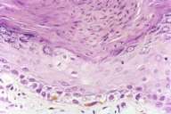
| Photomicrographs of the skin biopsy demonstrating coronoid lamellae. Low power (Fig. 2). Medium power (Fig. 3). | |

|

|
| Figure 4 | Figure 5 |
|---|---|
| High power (H&E, 100X) | |
Two skin biopsies taken from a seborrheic keratosis-like lesion (gluteal skin) and a typical porokeratosis lesion (forearm) revealed similar pathology, namely, multiple parakeratotic columns, the cornoid lamellae (Figs. 2-5). Beneath the cornoid lamellae, absence of granular layers and dyskeratotic cells were observed. The dermis showed superficial perivascular lymphohistiocytic infiltrate and melanophages.
Topical tretinoin 0.075 percent solution, 5-fluorouracil cream (5 %), and calcipotriol cream were tried on three adjacent areas for 6 weeks. Of the three, tretinoin seemed to be associated with the best clinical improvement. However, complete remission was not obtained.
Discussion
Porokeratosis is a disease characterized histologically by the presence of cornoid lamellae. Several well-established clinical types and variants have been described. The lesions of porokeratosis may be single or multiple. When multiple lesions are present, they may be generalized or localized (as observed in our patient). Although rare, porokeratosis has been reported to localize to certain anatomic areas, for example, vulva [1], or penis and scrotum [2]. The lesions in our patients were mostly clustered in the gluteal area.
Another peculiar feature noted in our patient is the resemblance of the lesions to seborrheic keratosis or verruca vulgaris. Hyperkeratotic forms of porokeratosis have been reported most frequently with the disseminated superficial actinic (DSAP) and the Mibelli types [3, 4, 5]. The lesions of our patient are not distributed in the sun-exposed areas and thus do not conform to DSAP type. The clinical morphology of the lesions observed in our patients is also not typical of porokeratosis of the Mibelli type, namely, lacking the peripheral ridges, and thus actually simulates seborrheic keratosis. Moreover, the lesions of porokeratosis of Mibelli tend to be few in numbers whereas the number of lesions in our patient was well over thirty. Although the morphology of the lesions reported herein does not simulate the hyperkeratotic form of porokeratosis of Mibelli [4], there are several similar features. The presence of multiple cornoid lamellae, the number of dyskeratotic cells at the base of the cornoid lamellae, which Jacyk and Esplin have described to be more extensive in the hyperkeratotic form than the classic porokeratosis of Mibelli [4], and the degree of dermal changes consisting of dense inflammatory infiltrate and dilated blood vessels are all thought to be unique to this entity [4].
The significance of recognizing porokeratosis lesions lies within the fact that cutaneous malignancy is known to arise within these lesions. The lesions that are especially at high risks are those which are large, of long duration, and the linear type of porokeratosis [6].
Several treatment modalities exist. There are reports citing the benefits obtained from topical and systemic retinoids [7, 8], topical vitamin D3 analogs [9], and 5-fluorouracil [10, 11]. Our patient seems to respond most favorably to topical tretinoin. Retinoids, especially when administered systemically, may be superior to other treatment modalities in light of their ability to improve cytologic atypia observed in some patients [8].
For the more recalcitrant lesions, surgical measures such as CO2 laser vaporization or resurfacing [12, 13] may be helpful. Surprisingly, even untreated lesions have disappeared following CO2 laser treatment delivered at remote sites [11].
References
1. Robinson JB, Im DD, Jockle G, Rosenshein NB. Vulvar porokeratosis: case report and review of the literature. Int J Gynecol Pathol 1999;18:169-1732. Neri I, Marzaduri S, Passarini B, Patrizi A. Genital porokeratosis of Mibelli. Genitourin Med 1995;71:410-411
3. Schuldenfrei JA, Fix LW. Disseminated superficial actinic porokeratosis with hyperkeratotic lesions. Cutis 1984;33:369-370
4. Jacyk WK, Esplin L. Hyperkeratotic form of porokeratosis of Mibelli. Int J Dermatol 1993;32:902-903
5. Jang KA, Choi JH, Sung KJ, Moon KC, Koh JK. The hyperkeratotic variant of disseminated superficial actinic porokeratosis (DSAP). Int J Dermatol 1999;38:204-206
6. Sasson M, Krain AD. Porokeratosis and cutaneous malignancy. A review. Dermatol Surg 1996;22:339-342
7. Danno K, Yamamoto M, Yokoo T, Ohta M, Ohno S. Etretinate treatment in disseminated porokeratosis. J Dermatol 1988;15:440-444
8. Goldman GD, Milstone LM. Generalized linear porokeratosis treated with etretinate. Arch Dermatol 1995;131:496-497
9. Harrison PV, Stollery N. Disseminated superficial actinic porokeratosis responding to calcipotriol (letter). Clin Exp Dermatol 1994;19:95
10. Shelley WB, Shelley ED. Disseminated superficial porokeratosis: rapid therapeutic response to 5-fluorouracil. Cutis 1983;32:139-140
11. McDonald SG, Peterka ES. Porokeratosis (Mibelli): treatment with topical 5-fluorouracil. J Am Acad Dermatol 1983;8:107-110
12. Hunziker T, Bayard W. Carbon dioxide laser in the treatment of porokeratosis. J Am Acad Dermatol 1987;16:625
13. Barnett JH. Linear porokeratosis: treatment with the carbon dioxide laser. J Am Acad Dermatol 1986;14:902-904
© 2005 Dermatology Online Journal

