Hyperpigmented burn scar improved with a fractionated 1550 nm non-ablative laser
Published Web Location
https://doi.org/10.5070/D396n5f8qnMain Content
Hyperpigmented burn scar improved with a fractionated 1550 nm non-ablative laser
Daniel Q Bach1 BA, Miki S Garcia2 MD, Daniel B Eisen2 MD
Dermatology Online Journal 18 (7): 12
1. Feinberg School of Medicine, Northwestern University Chicago, Illinois2. University of California Davis Medical System, Department of Dermatology, Sacramento, California
Abstract
Scars sustained following injury in patients with darker skin types can present a treatment challenge. These scars often hyperpigment and may remain refractory to first line treatments such as topical retinoids and hydroquinone. Additionally, more aggressive treatment interventions such as ablative resurfacing, chemical peels, and Q-switched laser therapy may actually worsen the pigmentation. We describe a 22-year female with a hyperpigmented scar and Fitzpatrick type IV skin that improved markedly following treatment with a fractionated erbium doped fiber laser. The improvement was maintained at least 1 year following the last procedure.
Case
A 22-year-old female presented for evaluation and treatment of her hyperpigmented scar on her right upper arm from a burn injury at work when boiling water spilled onto her. Prior treatment to the site included triamcinolone 0.1 percent cream for 2 months to help quell inflammation and irritation around the area. Hydroquinone 4 percent cream was subsequently applied twice daily in an attempt to help reduce the hyperpigmentation in the scar. No improvement in discoloration was seen after more than 4 months of treatment with the hydroquinone. The patient continued to practice proper sun protection to prevent further darkening of the scar site.
 |  |
| Figure 1 | Figure 2 |
|---|---|
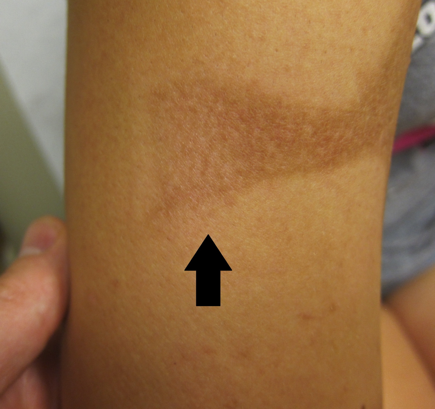
| Figure 1. Hyperpigmented burn scar on initial presentation prior to treatment with fractional non-ablative laser. Figure 2. Post-operative photo 1 month after treatment performed at test spot on right lateral edge of scar (arrow). | |
Upon presentation, the patient, skin phototype IV, revealed a large, approximately 8 by 6 centimeter, hyperpigmented, dark brown plaque on the upper right anterior arm with minimal evidence of textural changes including hypertrophy or atrophy (Figure 1).
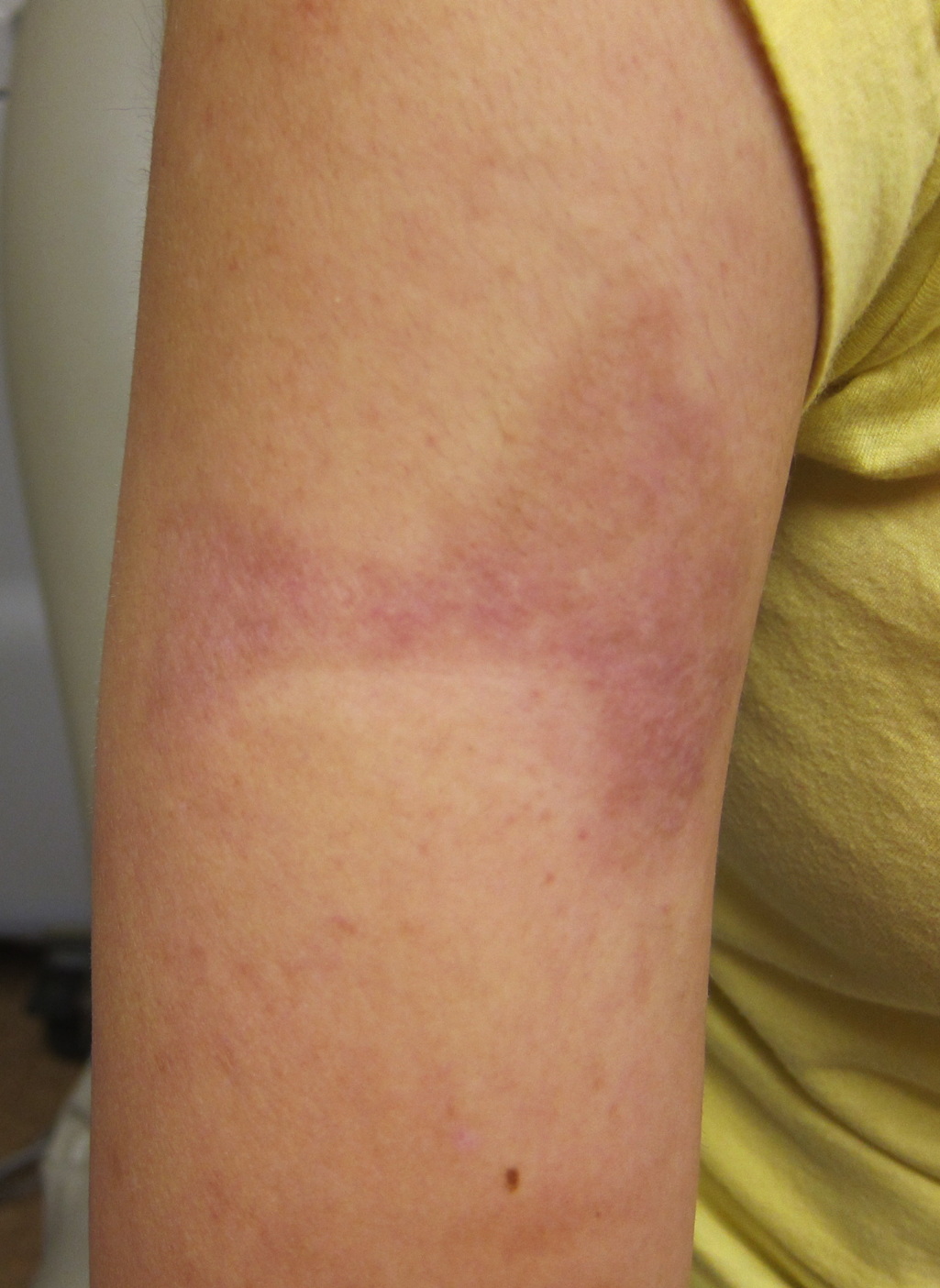 |
| Figure 3 |
|---|
| Figure 3. Post-operative photo following 3 full treatments of the entire scar area. |
Given previous failure of conventional topical therapies and patient desire, use of the fractional non-ablative laser 1550 nm wavelength (Fraxel re:store, Solta Medical Inc, Hayward, CA) was considered. A test spot was performed at the right lateral edge of the scar. A 15 mm treatment tip was used at 15 mJ, treatment level 6, with a Zimmer cooler on a setting of 6. The patient tolerated the treatment well and returned in one month for assessment of treatment efficacy. At the time she was noted to have significant lightening of pigmentation in the treated area (Figure 2). Subsequently, treatment of the entire scar was performed for a total of 3 additional treatments under similar parameters, except a decrease from 15 mJ to 13 mJ after the second treatment because of the patient report of mild tenderness during and 1 to 2 days directly after treatment. The patient, however, denied having any worsening pain or evidence of blistering.
Following 3 treatments and 1 test spot treatment, the hyperpigmentation of her scar site secondary to the water burn injury was markedly reduced (Figure 3). Further treatments were stopped with no evidence of re-pigmentation or textural changes being seen 1 year out following her last treatment. The patient was pleased with the treatment and final appearance.
Discussion
Fractional photothermolysis is a relatively new concept that was first introduced in 2004 as an alternative method to conventional ablative and non-ablative laser technologies [1]. The major advantage of fractional photothermolysis, in comparison to prior laser technologies, is its ability to target only a fraction of the skin. The thermal damage induced by the laser is confined to columns in the dermis termed microscopic thermal zones (MTZs) with the density and depth being adjusted as desired by varying the wavelength and pulse energy. Most importantly, this focal targeting leaves healthy skin around each MTZ intact; epithelial cells can more quickly migrate into and simulate collagen formation for effective wound healing. The effect is faster clinical recovery times, reduced side effects, and improved outcomes [1, 2].
Since its initial introduction, many indications have been reported for this new laser system including treatment of pigmented lesions, rhytides, facial dyschromias, actinic keratosis, and acne and surgical scars [3]. Recently, there have also been reports of the use of fractional lasers for resurfacing scars resulting from thermal burns [4, 5]. However, these scars were not hyperpigmented. Katz et al. reported improvement in an unscarred area with post inflammatory hyperpigmentation using similar parameters as for our patient [6].
Improvement of facial dyschromias is theorized to occur via a melanin shuttle [7]. Following laser induced thermal damage to the treated skin, dermal and epidermal components, including melanin, are eliminated through the overlying epidermis [1, 7, 8]. The improvement noted in our patient was likely the result of this mechanism. We hypothesize that the relatively low fluence used on our patient, as well as the large percentage of the skin surface undamaged (typical of fractional resurfacing) may have contributed to the lack of hyperpigmentation following the procedure.
Our case highlights the effectiveness and minimal side effects of the use of fractional non-ablative lasers to treat a hyperpigmented scar resulting from a burn injury. This represents one of the first reports for this indication. It is unknown whether hyperpigmented scars respond or carry risks related to treatment that are any different than other pigmentary disorders such as melasma, or post inflammatory hyperpigmentation occurring on unscarred skin. Fractional resurfacing may be a reasonable treatment for hyperpigmented scars that fail to respond to topical retinoids and hydroquinone. Further studies with larger numbers of patients will be necessary to gauge the true risks and benefits of this procedure, but our initial experience was quite positive.
References
1. Manstein, D., et al., Fractional photothermolysis: a new concept for cutaneous remodeling using microscopic patterns of thermal injury. Lasers Surg Med, 2004. 34(5): p. 426-38. [PubMed]2. Khan, M.H., et al., Intradermally focused infrared laser pulses: thermal effects at defined tissue depths. Lasers Surg Med, 2005. 36(4): p. 270-80. [PubMed]
3. Bogdan Allemann, I. and J. Kaufman, Fractional photothermolysis--an update. Lasers Med Sci, 2010. 25(1): p. 137-44. [PubMed]
4. Waibel, J. and K. Beer, Fractional laser resurfacing for thermal burns. J Drugs Dermatol, 2008. 7(1): p. 59-61. [PubMed]
5. Waibel, J. and K. Beer, Ablative fractional laser resurfacing for the treatment of a third-degree burn. J Drugs Dermatol, 2009. 8(3): p. 294-7. [PubMed]
6. Katz, T.M., et al., Fractional photothermolysis for the treatment of postinflammatory hyperpigmentation. Dermatol Surg, 2009. 35(11): p. 1844-8. [PubMed]
7. Hantash, B.M., et al., Laser-induced transepidermal elimination of dermal content by fractional photothermolysis. J Biomed Opt, 2006. 11(4): p. 041115. [PubMed]
8. Laubach, H.J., et al., Skin responses to fractional photothermolysis. Lasers Surg Med, 2006. 38(2): p. 142-9. [PubMed]
© 2012 Dermatology Online Journal