Pachyonychia congenita associated with median rhomboid glossitis
Published Web Location
https://doi.org/10.5070/D38nq996d7Main Content
Pachyonychia congenita associated with median rhomboid glossitis
Julie K Karen MD, Julie V Schaffer MD
Dermatology Online Journal 13 (1): 21
New York University Department of DermatologyAbstract
A 3-year-old girl presented with subungual hyperkeratosis and nail plates with increased transverse curvature, distal elevation, yellow-brown discoloration, and mild thickening. The changes, which affected all 20 nails, had developed during the first year of life. Mucocutaneous examination showed the presence of median rhomboid glossitis. The patient's mother had similar nail changes, which had been present since infancy as well as a focal plantar keratoderma and hyperhidrosis. The patient's clinical presentation and history were compatible with a diagnosis of pachyonychia congenita, a rare heritable disease that affects the nails, skin, oral and laryngeal mucosae, teeth, and hair. Dominant-negative mutations in four keratin genes (K6a, K6b, K16, and K17) lead to keratinocyte fragility and the resultant pachyonychia congenita phenotype. Successful targeted therapies are currently lacking for this oftentimes disabling disorder. Although oral manifestations are a common feature of PC, to our knowledge, this represents the first report of median rhomboid glossitis in association with PC.
Clinical synopsis
A 3-year-old girl presented to the Charles C. Harris Skin and Cancer Pavilion Pediatric Dermatology Clinic for the evaluation of asymptomatic nail changes that had developed during the first year of life. Her parents reported spontaneous shedding and then subsequent regrowth of the patient's right second fingernail at age 2 years. They did not know the duration of the painless lesion on the tongue and had not noticed any other mucocutaneous lesions. There was no history of natal or neonatal teeth, loss of primary teeth, acral blisters, plantar pain, or hoarseness. The patient had no known medical problems, and her growth and development have been normal. Her mother had similar nail abnormalities, which had developed during infancy and affected all 20 of her nails as well as a focal plantar keratoderma and hyperhidrosis. No other family members were affected.
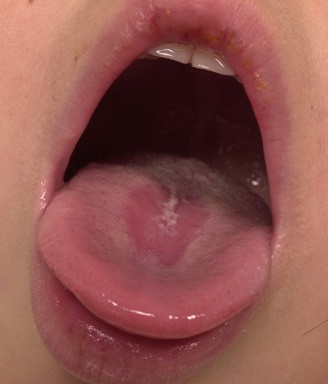
All 20 nails demonstrated subungual hyperkeratosis that was associated with distal elevation and increased transverse curvature of the nail plate, which leads to an omega appearance. The nail plates also were thick and yellow-brown in color. A sharply-demarcated, slightly-depressed, erythematous, smooth patch with a rhomboidal shape was present overlying the medial aspect of the dorsal surface of the tongue just anterior to the circumvallate papillae. No palmoplantar keratoderma, follicular keratoses, or cutaneous cysts were evident. The hair and teeth were normal, and there were no other oral mucosal lesions.
 |  |
| Figure 1 | Figure 2 |
|---|---|
Comment
Pachyonychia congenita (PC) represents a group of rare, autosomal dominant keratin disorders with characteristic nail findings and with the additional abnormalities of the palmoplantar skin, pilosebaceous apparatus, oral and laryngeal mucosae, teeth, and hair. Dominant-negative mutations producing aberrant keratin proteins that interfere with keratin filament assembly and lead to keratinocyte fragility are central to the pathogenesis of PC. The molecular bases of the two major clinical types of PC, PC-1 (Jadassohn-Lewandowski type) and PC-2 (Jackson-Lawler type), were elucidated in the 1990's. Defects in the genes encoding keratins 6a and 16 were found to underlie PC-1, whereas defects in keratins 6b and 17 were shown to underlie PC-2. To date, more than 80 mutations in these four keratin genes have been identified in PC families, the vast majority of which occur within the helix boundary motifs that flank the central helical rod domain [1]. Variants of PC-1 and PC-2 with delayed onset (PC tarda) represent exceptions that result from mutations elsewhere in the genes that encode keratins 16 and 17, respectively [2].
The hallmark of PC is hyperkeratosis of the nail bed, which leads to elevation, which is most pronounced distally, and increased transverse curvature of the nail plate [3]. This abnormality results in an omega or pincer nail. The nail plates are also discolored, thick, and friable, and they sometimes fail to reach the distal fingertip. All 20 nails are involved, although the findings are often most severe on thumbs, index fingers, and toes.
Additional features of PC, in order of decreasing prevalence, include: focal palmoplantar keratoderma (plantar, 90 %; palmar, 50 %); palmoplantar hyperhidrosis (75 %); oral leukokeratosis (50 %, more prominent in PC-1); follicular keratoses in sites of friction, such as the elbows, knees, and waistline (50 %); natal or neonatal teeth (PC-2, 50 %; PC-1, 0 %); cutaneous cysts (25 %, more common in PC-2); coarse or twisted hair (25 %, more common in PC-2); hoarseness due to laryngeal involvement (15 %); and corneal abnormalities (<5 %) [1]. A history of natal or neonatal teeth is highly suggestive of PC-2. Whereas epidermal inclusion cysts can be observed in both types of PC, the presence of steatocystomas or vellus hair cysts points to a diagnosis of PC-2. However, the typical post-pubertal onset limits this feature's utility as an early discriminating factor. Although the presence of pili torti also favors a diagnosis of PC-2, brittle, coarse hair has been observed in both types. The development of painful oral and nipple lesions during breastfeeding and copious production of ear wax were recently described as additional clinical manifestations of PC [1].
Quality of life assessments have noted that plantar pain is the most disabling feature of PC. The development of plantar keratoderma is typically delayed until the child begins to walk, and blisters often precede the hyperkeratosis. Exquisite plantar pain commonly results in an inability to walk without an ambulatory aid, which is an underreported morbidity of the disease [1].
Oral manifestations are frequently the earliest sign of PC and most commonly comprise leukokeratosis of the tongue and buccal mucosa. When the buccal mucosa is involved, accentuation along the bite line usually is noted. Clinically, these lesions may mimic oral candidiasis, white-sponge nevus, hairy tongue, or premalignant leukoplakia. However, malignant degeneration has not been reported [1]. Angular cheilitis, which is often associated with a candidal infection, also has also been described in association with PC [4].
To our knowledge, median rhomboid glossitis (MRG) has not previously been reported in association with PC. Median rhomboid glossitis appears as a shiny, rhomboidal or diamond-shaped, depapillated, erythematous patch on the medial aspect of the dorsal surface of the tongue just anterior to the circumvallate papillae [5]. A less common presentation that involves the paramedial area of the tongue has been termed atypical rhomboid glossitis [6]. Median rhomboid glossitis has been reported to occur in association with candidal cheilitis, and palatine kissing lesions may represent a sign of human immunodeficiency virus infection or other immunodeficiency states [7]. The lesion may be asymptomatic, as in our patient, or may be associated with a burning sensation [5]. Median rhomboid glossitis was once thought to represent a developmental anomaly that was related to persistence of an embryonic structure, the tuberculum impar. However, MRG is now considered to be a localized, chronic infection with Candida albicans. This belief is buttressed by reports of yeast and pseudohyphae in lesional scrapings, positive cultures, and rapid clearance with antifungal therapy [5]. Treatment of our patient with nystatin suspension for several weeks led to some improvement but not clearance of the MRG, which suggests that PC and candidiasis were both factors in its pathogenesis.
Like most genodermatoses, there are currently no specific treatments for PC [8]. Therapy is directed at the manifestations that are most troublesome to individual patients. Treatment of the nail alterations is challenging. Avulsion of distorted nails provides only temporary benefit, as regrowth is often associated with more severe distortion [3]. Ablation of the nail matrix has been reported to improve function and appearance in some patients [9]. Overnight occlusion with urea or salicylic acid pastes in conjunction with mechanical paring has been variably successful. Systemic retinoid therapy sometimes improves oral leukokeratosis and follicular keratoses but has little effect on keratoderma or nail dystrophy [3]. Twice daily application of 20 percent aluminum chloride solution followed by 10 percent salicylic acid ointment has been reported to alleviate plantar hyperhidrosis and painful ambulation. [10] Plantar injections of botulinum toxin type A after intravenous regional anesthesia has led to relief of pain and hyperhidrosis in three patients with severe plantar involvement. The duration of improvement ranged from 6 weeks to 6 months, and tachyphylaxis did not occur in individuals who underwent repeated treatments [11].
Although the ultimate goal of targeted gene correction is still impractical, gene therapy aimed at suppressing production of the abnormal protein represents a plausible option for diseases caused by dominant-negative keratin mutations. Considering the accessibility of the skin, advancements in delivery systems, and the availability of RNA reduction agents, such as antisense oligonucleotides, triplex-forming oligonucleotides, and small interfering RNA, gene silencing approaches to the treatment of PC may be feasible in the near future [12].
References
1. Leachman SA, et al. Clinical and pathological features of pachyonychia congenita. J Investig Dermatol Symp Proc 2005;10:32. Paller AG, et al. Pachyonychia congenita tarda. Arch Dermatol 1991;127:701
3. Dahl PR, et al. Jadassohn-Lewandowski syndrome (pachyonychia congenita). Semin Dermatol 1995;14:129
4. Su WP, et al. Pachyonychia congenita: a clinical study of 12 cases and review of the literature. Pediatr Dermatol 1990;7:33
5. Bae GY, et al. A case of median rhomboid glossitis. J Dermatol 2003;30:423
6. Lago-Mendez L, et al. Rhomboid glossitis in atypical location: case report and differential diagnosis. Med Oral Patol Oral Cir Bucal 2005;10:123
7. Brown RS, Krakow AM. Median rhomboid glossitis and a "kissing" lesion of the palate. Oral Surg Oral Med Oral Pathol Oral Radiol Endod 1996;82:472
8. Kaspar RL. Challenges in developing therapies for rare diseases including pachyonychia congenita. J Investig Dermatol Symp Proc 2005;10:62
9. Thomsen RJ, et al. Pachyonychia congenita: surgical management of the nail changes. J Dermatol Surg Oncol 1982;8:24
10. Takayama M, et al. Alleviation of the plantar discomfort caused by pachyonychia congenita with topical applications of aluminum chloride and salicylic acid ointments. J Dermatol 2005;211:302
11. Swartling C, Vahlquist A. Treatment of pachyonychia congenital with plantar injections of botulinum toxin. Br J Dermatol 2006;154:763
12. Lewin AS, et al. Gene therapy for autosomal dominant disorders of keratin. J Investig Dermatol Symp Proc 2005;10:47
© 2007 Dermatology Online Journal