Atypical cutaneous leishmaniasis resembling eczema on the foot
Published Web Location
https://doi.org/10.5070/D385p9d1stMain Content
Atypical cutaneous leishmaniasis resembling eczema on the foot
A Manzur1, and UA Butt2
Dermatology Online Journal 12 (3): 18
1.Department of Dermatology PAF Hospital Sargodha, Pakistan. aamirderm@hotmail.com2. Department of Dermatology Combined Military Hospital Muzaffarabad, Pakistan.
Abstract
Cutaneous leishmaniasis (CL) typically presents as ulcerated or crusted nodules and plaques. Eczematous morphology of CL lesions is unusual. We report a case of CL that presented as a large eczematous plaque with no clinically obvious primary lesion.
Localized cutaneous leishmaniasis (CL) typically presents as papules, nodules, plaques or noduloulcerative lesions. The unusual clinical presentations for localized CL are reported occasionally and include hyperkeratotic psoriasiform lesions, eczematoid lesions, zosteriform pattern, warty lesions, erysipeloid, and acneiform lesions [1, 2]. The incidence of eczematoid form of CL is reported to be 2.3 percent from a study in Iran [2].
Clinical synopsis
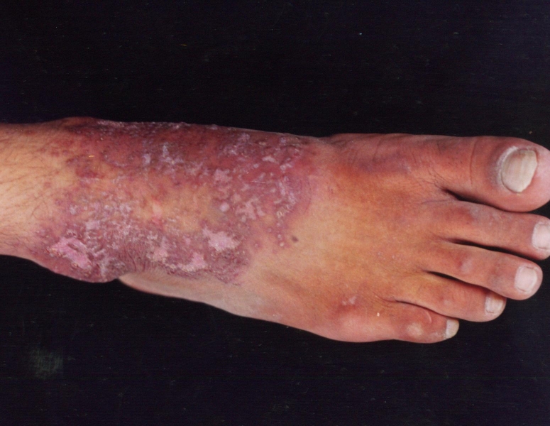 |
| Figure 1 |
|---|
A 21-year-old man presented with a 3-week history of a pruritic exuding lesion on the right foot. It started as a small insect-bite-like lesion. The patient denied history of trauma, allergic disorders, atopic predisposition, or drug intake. On examination, there was a 14 × 10 cm crusted plaque involving anterior surface of ankle and dorsum of the right foot. The plaque had dirty-brown crust and multiple papulopustules exuding a thick yellow discharge. Right inguinal lymph nodes were enlarged and mildly tender. There was no palpable lymphatic cord. Routine laboratory investigations were normal including a negative HIV serology. Pus culture grew Staphylococcus aureus sensitive to cephalexin. The clinical picture was consistent with acute infected eczema. Various treatment attempts with antibiotics and steroids failed to heal the lesion, so a skin biopsy was done. By this time the lesion had dried up considerably (Fig. 1). Histopathology revealed a dense dermal infiltrate of macrophages, lymphocytes, plasma cells, and numerous Leishmania amastigotes (both intra- and extra-cellular). The epidermis showed hyperkeratosis and acanthosis, but no amastigotes were seen within the epidermis. The patient responded to parentral meglumine antimonate (20 mg/kg body wt), with complete healing of lesion after a 12-day treatment.
Discussion
The clinical picture in leishmaniasis depends not only on the infecting Leishmania species, but also on the host immune response, which is largely mediated through cellular immunity. Other factors that affect the clinical picture include the number of parasites inoculated, site of inoculation, nutritional status of the host and nature of the last non-blood meal of the vector [3]. Factors such as a non-indigenous individual [2], HIV infection [4], use of oral steroids [5], old age [6], and even wound contamination with inorganic materials [7] can alter the clinical picture of CL. Similarly, the unusual morphology of CL lesions reported in atopics is likely associated with an imbalance between TH1 and TH2 cells [8] or decreased chemotaxis, and persistent pruritus [9].
The precise pathogenesis of the eczematous appearance of the localized CL has been poorly documented. However, one factor could be the epidermal invasion by parasites causing an intense cell-mediated immune response leading to severe inflammatory and eczematous changes [10].
In our patient the uniform appearance and large size of the lesion was in itself very unusual, because there was no primary plaque or nodule. The lesion size cannot be attributed to the satellite papules because the biopsy taken from the periphery had an abundance of amastigotes; satellite papules are usually negative for amastigotes [1]. For the same reason, the lesion morphology cannot solely be ascribed to secondary bacterial infection. Our patient was an otherwise healthy, HIV-negative, young adult with no history of atopy or any other skin or systemic disease to account for the unusual clinical picture. Whether the atypical morphology resulted from multiple sandfly bites, from a specific immune response or lack of response, from an atypical Leishmania strain, or from non-indigenous status of the patient is not clear.
The growing phenomenon of international tourism and military operations in highly endemic areas stresses the importance of a high clinical suspicion in patients residing or having visited these regions. In these circumstances it is recommended that CL should be included in the differential diagnosis of recalcitrant eczematous eruptions. Timely diagnosis of CL can avoid complications and help institute an early and effective treatment.
References
1. Kubba R, AI-Gindan Y, EI-Hassan AM, Omer AH. Clinical diagnosis of cutaneous leishmaniasis (oriental sore). J Am Acad Dermatol 1987; 16: 1183-1189. PubMed.2. Momeni AZ, Aminjavaheri M. Clinical picture of cutaneous leishmaniasis in Isfahan, Iran. Int J Dermatol 1994; 33: 260-265. PubMed
3. Farah FS, Klaus SN, Frankenburg S, et al. Protozoan and helminth infections. In: Dermatology in General Medicine, Vol ll, 4th Ed. New York: McGraw-Hill; 1993; 2772-2777.
4. Puig L, Pradinaud R. Leishmania and HIV co-infection: dermatological manifestations. Ann Trop Med Parasitol 2003; 97 Suppl 1: 107-114. PubMed
5. Motta AC, Arruda D, Souza CS, Foss NT. Disseminated mucocutaneous leishmaniasis resulting from chronic use of corticosteroid. Int J Dermatol 2003; 42(9): 703-706. PubMed
6. Salmanpour R, Handjani F, Zerehsaz F, Ardehali S, Panjehshahin MR. Erysipeloid leishmaniasis: an unusual clinical presentation. Eur J Dermatol 1999; 9(6): 458-459. PubMed
7. Convit J, Ulrich M, Perez M, Hung J, Castillo J, Rojas H, Viquez A, Araya LN, Lima HD. Atypical cutaneous leishmaniasis in Central America: possible interaction between infectious and environmental elements. Trans R Soc Trop Med Hyg 2005; 99(1): 13-17. PubMed
8. Guarneri C, Vaccaro M, Cannavo SP, Borgia F, Guarneri. Erythematous-edematous-infiltrative plaque on the face: cutaneous angio-lupoid leishmaniasis. Eur J Dermatol 2002; 12(6): 597-599. PubMed
9. Kubeyinje E P, Belagavi C S. Cutaneous leishmaniasis occurring with atopic eczema: report of three cases. East Afr Med J 2000; 77(10): 572-573. PubMed
10. Uzun S, Acar MA, Uslular C, Kavukcu H, Aksungur VL, Culha G, Gurel MS, Memisoglu HR. Uncommon presentation of cutaneous leishmaniasis as eczema-like eruption. J Eur Acad Dermatol Venereol 1999; 12(3): 266-268. PubMed
© 2006 Dermatology Online Journal