Disseminated primary HSV-2 infection of the face
Published Web Location
https://doi.org/10.5070/D38206c7q6Main Content
Letter: Disseminated primary HSV-2 infection of the face
Elie Maalouf1 MD, Roy Moutran2 MD, Ismael Maatouk1 MD
Dermatology Online Journal 18 (6): 15
1. Hotel-Dieu de France, Achrafieh, Beirut, Lebanon2. Mount-Lebanon Hospital, Hazmieh, Beirut, Lebanon
Abstract
We report the case of a 44-year-old, heterosexual, man, who presented for lesions of the face that appeared 3 days earlier; the eruption was associated with a burning sensation. He had sexual intercourse 12 days prior to presentation with a new partner. On clinical examination, there were confluent vesicules and a few pustules localized on the cheeks, forehead, nose, mouth, and ears. A swab for immunofluorescence (IF) came back as positive for HSV-2. The patient was treated with oral acyclovir. The lesions were healed when he was seen for follow-up 1 week later. The virus responsible for herpes is a double-stranded DNA virus named Herpes simplex virus (HSV). The virus generally enters damaged epithelium or mucosal surfaces, secondary to abrasions or trauma. Most primary orolabial infections occur during childhood as herpetic gingivostomatitis. However, there are forms that could be more atypical. The spread of the virus was probably promoted by shaving the beard. In immunocompromised patients or those with skin barrier disorders, HSV infection tends to disseminate and is accompanied by visceral involvement. Hence, the need to detect a state of immunodepression (including AIDS) in any patient with diffuse herpes infection. Three oral antiviral agents are commonly used: acyclovir, famciclovir, and valaciclovir.
Case
A 44-year-old, heterosexual, man came in consultation to the dermatology department for lesions of the face that appeared 3 days earlier. His past medical history consisted of a nephrectomy and urinary tract infections. The patient had no history of orolabial herpes.
The patient reported the appearance of facial skin lesions 3 days prior to his office visit and he noted a burning sensation. He also noted mild photophobia with tearing. The patient used to shave his beard 1-2 times a week. While reviewing his recent activities, he admitted having had sexual intercourse 12 days prior with a new partner.
 |  |
| Figure 1 | Figure 2 |
|---|---|
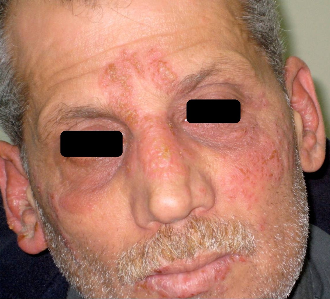
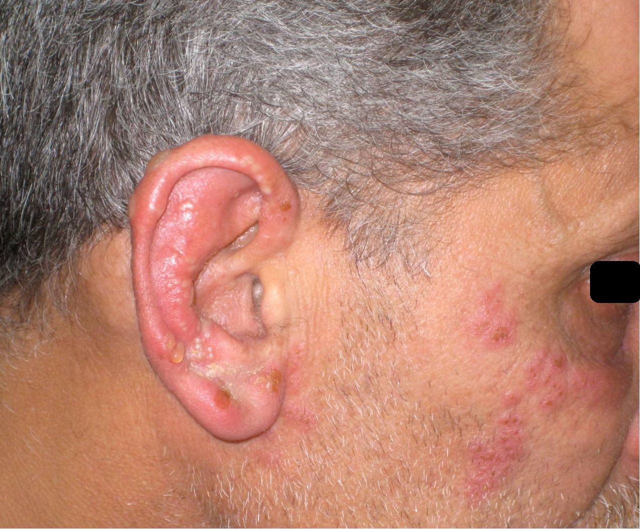
| Figure 1. Vesicules on the cheeks, forehead, nose and mouth Figure 2. Vesicules on the right cheek and right ear | |
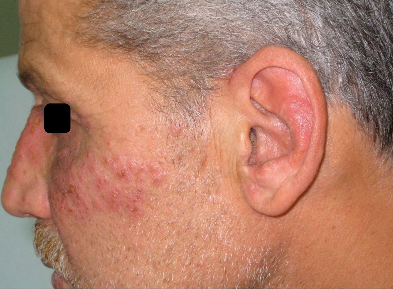
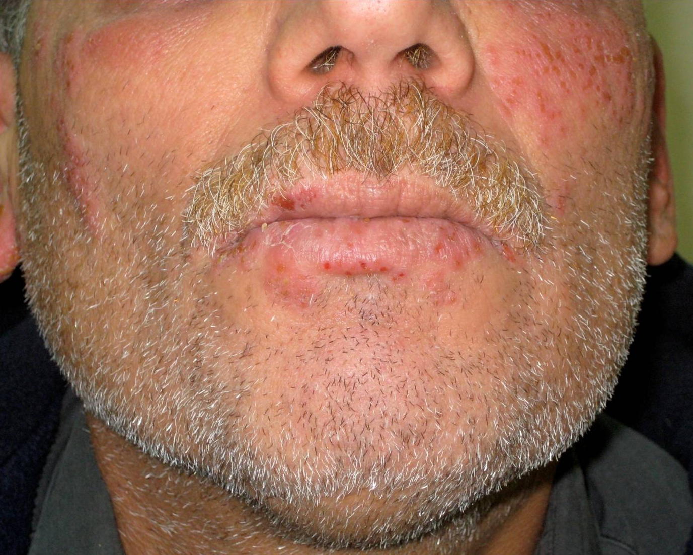
On clinical examination, there were confluent vesicules, a few pustules, and edematous eyelids. The lesions were localized on the cheeks, forehead, nose, mouth, ears, and lips (Figures 1, 2, 3, and 4). No intraoral lesions were detected. There were no signs of facial eczema or other dermatitis.
 |  |
| Figure 3 | Figure 4 |
|---|---|
| Figure 3. Vesicular lesions on the left cheek and left ear Figure 4. Vesicules around the lips. No intra-oral dermatitis. | |
Because mucocutaneous herpes was suspected, a swab for IF was performed, testing positive for Herpes simplex virus 2 (HSV-2). Ophthalmologic examination did not reveal ocular involvement.
The patient was treated with acyclovir 200 mg 5x/day for one week. The lesions were healed when he was seen for follow-up one week later. The assessment for immunodeficiency, liver function tests, and viral serology were normal.
Discussion
The virus responsible for herpes is a double-stranded DNA virus of 150 to 200 nm named Herpes simplex virus (HSV). Two subtypes exist: HSV-1 and HSV-2, differentiated by their structural criteria and epidemiology. The human species is the only reservoir of the virus and transmission is interhuman by direct contact. Women are more often infected, and infection rates increase with age in men and women [1].
Usually, the virus enters damaged epithelium or mucosal surfaces, secondary to abrasions or trauma [2]. On the other hand, keratinocytes and dendritic cells are known to have receptors that allow the entry of HSV.
Most primary oro-labial infections occur during childhood. The infection is symptomatic in only 5 to 10 percent of children, presenting as herpetic gingivostomatitis, which lasts for 14 to 24 days [3, 4]. However, there are forms that could be more atypical, as was the case of our patient with diffuse cutaneous primary infection on the face in a 44-year-old male. The spread of the virus was probably promoted by shaving the beard. The alteration of the barrier function of skin is responsible for the dissemination of HSV.
Herpes gladiatorum [5] is a particularly diffuse form, related to massive contamination by the herpes virus as a result of close bodily contact, injuries, and abrasions caused by the practice of a contact sport. In these cases, the right side of the face is the most frequently involved because most wrestlers are right-handed [6].
In immunocompromised patients (hematopoietic disorders, after bone marrow or solid organ transplantation, patients on immunosuppressive therapy), newborns, pregnant women, or patients with skin barrier disorders (atopic dermatitis, Darier disease), HSV infection may become disseminated and is accompanied by visceral involvement, sometimes fatal. This form is quite rare in immunocompetent individuals [7]. Hence, it is necessary to investigate for immunodepression in any patient with diffuse herpes infection. The work up was negative in our patient. Similarly, HIV status was negative. This must be systematically checked since the co-infection HSV/HIV occurs in 50 to 95 percent according to studies [8]. Currently, three oral antiviral agents are commonly used in the treatment of HSV outbreaks: acyclovir, famciclovir, and valaciclovir [9].
References
1. Keller EC, Tomecki KJ. Cutaneous infections and infestations: new therapies. J Clin Aesthet Dermatol. 2011;4(12):18-24. [PubMed]2. Cunningham AL, Diefenbach RJ, Miranda-Saksena M, et al. The cycle of human herpes simplex virus infection: virus transport and immune control. J Infect Dis 2006; 194 (Suppl. 1): S11-S18. [PubMed]
3. Whitley RJ. Herpes simplex virus infections. In: Glaser R, Jones JF, editors. Herpesvirus infections. New York: Marcel Dekker,1994:1-58.
4. Nikkels AF, Pièrard GE. Treatment of mucocutaneous presentations of herpes simplex virus infections. Am J Clin Dermatol. 2002;3(7):475-87. [PubMed]
5. Belongia EA, Goodman JL, Holland EJ, et al. An outbreak of herpes gladiatorum at a high-school wrestling camp. N Engl J Med. 1991;325(13):906-10. [PubMed]
6. Anderson BJ. Managing herpes gladiatorum outbreaks in competitive wrestling: the 2007 Minnesota experience. Curr Sports Med Rep. 2008;7(6):323-7. [PubMed]
7. Watanabe D, Kuhara T, Ishida N, et al. Disseminated mucocutaneous herpes simplex virus infection in an immunocompetent woman. Int J STD AIDS. 2010;21(3):213-4. [PubMed]
8. Schacker T. The role of HSV in the transmission and progression of HIV. Herpes. 2001;8:46-8. [PubMed]
9. Patel AR, Romanelli P, Roberts B, Kirsner RS. Herpes simplex virus: a histopathologic study of the depth of herpetic wounds. Int J Dermatol. 2009;48(1):36-40. [PubMed]
© 2012 Dermatology Online Journal