Detection of high-risk human papillomavirus type 16/18 in cutaneous warts in immunocompetent patients, using polymerase chain reaction
Published Web Location
https://doi.org/10.5070/D37g5565r2Main Content
Detection of high-risk human papillomavirus type 16/18 in cutaneous warts in immunocompetent patients, using polymerase chain
reaction
R Payal MD1 S Gupta R Aggarwal MD DNB, S Handa MD2, BD Radotra MD PhD3, SK Arora PhD MNAMS
Dermatology Online Journal 12 (6): 1
Department of Immunopathology, Pathology1, Dermatology2 and histopathology3, Postgraduate Institute of Medical Education and Research, Chandigarh, IndiaAbstract
Cutaneous warts are caused by human papillomavirus (HPV). Prevalence studies of the types of HPV present in cutaneous warts have been carried out more frequently in immunosuppressed patients. The present study was designed to study the association of high-risk HPV in cutaneous warts of immunocompetent patients. A total of 45 cases of cutaneous warts from various sites in immunocompetent subjects were analyzed for HPV. Samples included both archival material i.e., paraffin embedded and fresh tissue. Highly sensitive and comprehensive polymerase chain reaction (PCR) methodology for detection of HPV of high oncogenic potential, HPV 16/18, was employed. Human papillomavirus 16 was detected in 3 (6.6 %) patients. None of the lesions demonstrated HPV 18. None of the cutaneous warts demonstrated histopathological features associated with dysplasia or neoplasia. The identification of HPV 16 in cutaneous warts, which are benign proliferations of the skin, further expands the spectrum of HPV-linked lesions. It remains of critical interest to determine whether these types are specifically associated with the development of malignant lesions analogous to those seen in anogenital cancer.
Introduction
Human papillomavirus (HPV) is the most ubiquitous of the human viruses. Over 100 HPV types have been identified. A majority of HPV infect the skin of normal as well as immunocompromised individuals. It appears that most HPV viruses establish a latent infection of the skin as normal flora residing in hair follicles. A minority of HPV are associated with cutaneous warts and mucosal condylomata [1]. Warts or verrucae are benign proliferations of the skin that take several forms, common warts (verrucae vulgaris), flat warts (verruca plana), plantar warts, mosaic warts and butcher's warts. Human papillomavirus may have a role in promoting proliferative lesions of the skin, although the site of active infection and mode of transmission is unknown.
Human papillomavirus is an epitheliotrophic virus and is inoculated into the viable epidermis through defects in the epithelium. Only a few types of HPV with oncogenic potential (low and intermediate) have been recognized in cutaneous warts. The more frequent HPV found in lesions of cutaneous common warts in the general population are HPV types 2, 57, 27, 4 and 1 [2, 3]. There are a paucity of data on the association of mucous membrane HPV of high oncogenic potential with cutaneous warts. The present study aims to analyze the association of HPV types 16 and 18 in cutaneous warts of immunocompetent patients.
Material and methods
Forty-five cases of clinically diagnosed cutaneous warts with subsequently histologically proven verrucae were recruited from January to December 2004. Lesions were considered as common warts if the lesions presented with compact hyperkeratosis, acanthosis, papillomatosis, hypergranulosis, elongated and flattened dermal papillae bent inwards towards the centre of the lesion, and enlargement of the capillaries (Fig. 1). Fresh tissue was available in 10 cases. In the other 35 cases, DNA was extracted from paraffin embedded tissue. Cutaneous warts were excised under aseptic conditions and local anaesthesia. A part of the tissue was stored at 4°C in phosphate-buffered saline (pH 7.4). The rest was preserved in 10 percent buffered formalin for histopathological examination. Ten cases of non-viral dermatological conditions were included as negative controls.
DNA extraction
The tissue specimen was teased and suspended in 500µl of lysis buffer containing 1 percent SDS and 0.01 percent proteinase K in Tris-EDTA (TE) buffer (pH 8.0) and incubated at 55 °C overnight. Extraction was done by phenol-chloroform- isoamyl alcohol mixture. DNA was precipitated with ice chilled isopropanol. DNA was pelleted out next day and resuspended in TE buffer. From the paraffin embedded tissue blocks, 5µm sections were cut and deparaffinized with organic solvent and digested with proteinase K solution [4]. All the other steps were the same as for the fresh tissue. DNA was quantitated spectrophotometrically. PCR for β-actin gene was done for each sample as an internal control.
Polymerase chain reaction (PCR) for HPV
All samples were subjected to PCR using primers specific for consensus sequence spanning the E6 open reading frame of high risk HPV type 16, 18, 31, 33 [5]. The sequence of forward primer was 5'-TGTCAAAAACCGTTGTGTCC-3' and that of reverse primer 5'-GAGCTGTCGCTTAATTGCTC-3'. Positive samples were subjected to PCR using type specific primers for HPV types 16 and 18 [6]. Forward primer for HPV 16 being 5'-ATTAGTGAGTATAGACATTA-3' and that of reverse primer was 5'-GGCTTTTGACAGTTAATACA-3'. The forward and reverse sequence of HPV type 18 specific primers was 5'-ACTATGGCGCGCTTTGAGGA-3' and 5'-GGTTTCTGGCACCGCAGGCA-3', respectively. The amplified gene fragments were of 109 bp and 334 bp for HPV 16 and 18, respectively, and were visualized on 2 percent agarose gels
Observations and results
Forty-five patients were recruited in the study. The male:female ratio was 4:1. The majority of patients (57 %) were in the age group of 21-40 years. Mean age was 35 years (range 14-76). Most of the verrucae were present on the exposed parts of the body i.e., hand 17(37.8%), foot 9 (20%), arm 6(13.3%), scalp 6 (13.3%) and face 5(11%). In one case verrucae were present on chest and abdominal wall. None of the cutaneous warts showed histopathological features associated with dysplasia or neoplasia. The duration of cutaneous warts among patients varied from 2 months to 2 years but was less than 1 year in 31 (68%) patients.
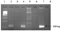
High risk oncogenic HPV type 16 was detected in 3 (6.6%) patients (Fig. 2). The lesions were positive with consensus primer for HPV types 16, 18, 31 and 33. These were confirmed using type specific primers for type 16 and 18. None of the lesion demonstrated HPV type 18. All the samples negative for high risk HPV were positive for β-actin primers, excluding failure of HPV amplification due to problems such as lack of DNA, inappropriate storage or technical problems. All 3 patients were aged less than 35 years. The lesions were present on the exposed parts of the body i.e., arm, hand and neck. None of the controls were positive for HPV.
Discussion
Warts are widespread in the worldwide population, although, the exact frequency is unknown. Human papillomavirus of low risk subtypes 2, 27, 57, 4, and 1 are known to be associated with cutaneous warts. Epidemiology as well as morphology of common warts is closely linked to the virus type. Prevalence studies on types of HPV present in cutaneous warts have been carried out more frequently in immunosuppressed patients. Multiple skin disorders such as warts, hyperkeratosis, keratoacanthomas, and other skin malignancies are more commonly seen in immunosuppressed patients, especially transplant patients. Studies regarding detection of HPV in skin warts using various techniques including PCR have revealed quite heterogenous results. Many primer pairs either degenerate or specific ones have been used by several authors with variable results regarding the percentage of HPV detection in cutaneous warts of immunocompetent and immunosuppressed patients. It is possible that diversity of results in published data may be due to variations among the prevalent HPV types in cutaneous warts as well as differences in the population studied.
Rubben et al. demonstrated that HPV types 2, 27, and 57 induced common warts at non-genital sites. Harvard et al. [7] detected mucous membrane HPV DNA by using a combination of primers by PCR and found a prevalence of 27.4 percent in immunosuppressed patients. Soler C et al. [8] suggested that HPV lose their specificity for mucosa and cutaneous sites in immunosuppressed patients. Chen et al. [9] detected HPV type 16 in 2 percent and type 18 in 8 percent respectively by using Southern blot hybridization technique. In the index study HPV 16 was detected in 6.6 percent of cases of cutaneous verrucae. Our data may be underestimating the true prevalence, because DNA was obtained in a majority of the cases from paraffin embedded tissue, which yields suboptimal results compared with fresh frozen tissue [10]. The reason we chose to use two primers set (i.e., consensus as well as type specific) was to increase the specificity of the test.
Cutaneous HPV types in the general population are predominantly associated with benign viral warts but their role in non-melanoma skin cancer has recently been postulated. The association of viral warts and skin cancer has been suspected in renal transplant patients for a long time [11]. Renal transplant patients have a well documented 50-100 fold increased risk of cutaneous squamous cell carcinoma [12]. The cumulative incidence of skin cancer is 27-44 percent after 10-25 years of immunosuppression [13]. With improved PCR techniques the association of HPV in non-melanoma skin cancer has been documented lately, although to a lesser extent. No particular HPV type has yet emerged as predominant.
Although high-risk HPV type 16 has been demonstrated in cutaneous warts, as in the present study, it is unknown what percentage may progress to develop neoplastic lesions. HPV 16 may remain latent for a prolonged period of time. It has also been suggested that HPV alone may not lead to development of skin cancer but this may require other additional factors such as sunlight (ultraviolet light) to lead to neoplastic diseases. Patients harboring high-risk HPV may progress to invasive squamous cell carcinoma after a long latency period of 20-50 years.
It will require large cross-sectional studies and long-term follow up, to confirm the association of different HPV types and non-melanoma skin cancer in immunocompetent patients. The diagnosis of HPV infection is an evolving field. DNA testing has greatly expanded the options available for the detection and study of HPV disease. So it remains of critical interest to determine which HPV types are specifically associated with the development of malignant cutaneous lesions analogous to those seen in anogenital cancers.
References
1. Jenson AB, Geyer S, Sundberg JP, Ghim S. Human Papillomavirus and skin cancer. J Investig Dermatol Symp Proc 2001; 6:203-6.2. Rubben A, Krones R, Schwetschenau B, Grussendorf-Conen E-1. Common warts from immunocompetent patients show the same distribution of human papillomavirus types as common warts from immunocompromised patients. Br J Dermatol 1993; 128:264-70.
3. Chan SY, Chen SH, Egawa K et al. Phylogenetic analysis of the human papillomavirus type 2 (HPV-2), HPV-27 and HPV-57 group, which is associated with common warts. Virology 1997; 239:296-302.
4. Shibata DK, Atheim N, Martin W. Detection of human papillomavirus in paraffin embedded tissue using the polymerase chain reaction. J Exp Med 1988; 167:225-230.
5. Oh YL, Shin KJ, Han J, Kim DS. Significance of high-risk human papillomavirus detection by polymerase chain reaction in primary cervical cancer screening. Cytopathol 2001; 12:75-83.
6. Miller CS, Zeuss MS, White DK. Detection of HPV DNA in oral carcinoma using polymerase chain reaction together with in situ hybridization. Oral Surg Oral Med Oral Pathol 1994; 77:480-6.
7. Harvard CA, Spink PJ, Surentheran T et al. Degenerate and nested PCR: a highly sensitive and specific method for detection of human papillomavirus infection in cutaneous warts. J Clin Microbiol 1999; 37:3545-3555.
8. Soler C, Allibert P, Chardounet V et al. Detection of human papillomavirus types 6, 11, 16 and 18 in mucosal and cutaneous lesions by the multiplex polymerase chain reaction. J Virol Methods 1995; 350:143-158.
9. Chen SL, Tsao YP, Lee JW et al. Characterisation and analysis of human papilloma viruses of skin wart. Arch Dermatol Res 1993; 285: 460-465.
10. Studniberg HM, Weller P. PUVA, UVB, psoriasis and non-melanoma skin cancer. J Am Acad Dermatol 1993; 29:1013-22.
11. Walder BK, Robertson MR, Jeremy D. Skin cancer and immunosuppression. Lancet 1971; ii: 1282-3.
12. Birkeland SA, Storm HH, Lamm LV et al. Cancer risk after renal transplantation in the Nordic countries, 1964-1986. Int J Cancer 1995; 60:183-9.
13. Hardie IR, Strong RW, Hartely LC et al. Skin cancer in Caucasian renal allograft recipients living in a subtropical climate. Surgery 1989; 87:177-83.
© 2006 Dermatology Online Journal


