Erythema elevatum diutinum
Published Web Location
https://doi.org/10.5070/D377s3f4bhMain Content
Erythema elevatum diutinum
Rachel Farley-Loftus MD, Chicky Dadlani MD, Nadia Wang MD, Karla Rosenman MD, Hideko Kamino MD, Steven Prystowsky MD, Andrew
Franks Jr MD, Miriam K Pomeranz MD
Dermatology Online Journal 14 (10): 13
Department of Dermatology, New York UniversityAbstract
A 64-year-old woman presented with a one-and-one-half year history of an enlarging, red-brown, firm plaque on the left thigh, with numerous, scattered, indurated, hyperpigmented patches on the lower extremities. Histopathologic examination of the largest plaque confirmed the diagnosis of erythema elevatum diutinum, which is a rare form of leukocytoclastic vasculitis that is associated with many disease entities, which include human immunodeficiency virus infection, malignant conditions, hematologic abnormalities, chronic infection, and autoimmune and connective-tissue disorders. The treatment of choice is dapsone; however, several other treatment modalities have been reported to be of benefit.
 |  |
| Figure 1 | Figure 2 |
|---|
History
A 64-year-old woman presented to the Dermatology Clinic of Bellevue Hospital Center in August, 2007, with an enlarging brown plaque on her left thigh The lesion initially presented as a firm, purple nodule, which began to enlarge over several months Several, scattered, hyperpigmented, firm plaques developed over the anterior and lateral surfaces of both legs She reported arthralgias and arthritis but denied a history of Raynaud phenomenon, dysphagia, dyspnea, fever, chills, or other systemic symptoms Her dermatologist performed a skin biopsy of the left thigh in November, 2006 At the same time, the patient also developed hypertension and rheumatoid arthritis, which was initially treated with prednisone and methotrexate Her regimen has since been changed to methotrexate and leflunomide, with stabilization of her joint disease.
Prior to her presentation to Bellevue Hospital Center, the skin lesions had been unresponsive to topical therapy with clobetasol cream, Dovonex, and pimecrolimus cream A second skin biopsy of the largest plaque on the left thigh was performed at Bellevue Hospital Center in October, 2007.
Physical Examination
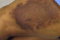
A large, brown, firm, indurated plaque with a cobblestone surface was present on the lateral aspect of the left thigh. Multiple, ill-defined, hyperpigmented, firm, indurated, nontender plaques were scattered over both lower extremities. No lesions were present on the trunk, face, or upper extremities. Lymphadenopathy was not detected.
Lab
A complete blood count, chemistry profile and liver function tests were normal. A Lyme Western blot was negative. Erythrocyte sedimentation rate was 27 mm/hr and rheumatoid factor was 134 IU/mL. Anti-nuclear antibody was negative. Quantitative IgA and IgE were normal; IgG was elevated at 1910 mg/dL. Serum protein electrophoresis showed diffuse hypergammaglobulinemia with no abnormal bands. Perinuclear-staining antineutrophil cytoplasmic antibodies (p-ANCA) was 15 U/mL (normal range<6). Glucose 6-phosphate dehydrogenase was zero. A chest radiograph showed no abnormalities.
Histopathology
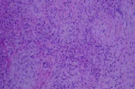
In the dermis and subcutaneous fat, there is a perivascular and interstitial, mixed-cell infiltrate composed of neutrophils with nuclear dust, lymphocytes, eosinophils, and some plasma cells. There are neutrophils and fibrin deposits in the walls of small blood vessels. There is fibrosis of the dermis and subcutaneous fat that is arranged as intersecting fascicles and concentric layers around the blood vessels. There are numerous, finely vacuolated histiocytes in the papillary and upper reticular dermis.
Comment
Erythema elevatum diutinum (EED) was initially described in elderly men [1] and in younger women with rheumatologic disease [2]. The two types were included together when this entity was reviewed and given its current name in 1894 [3, 4].
Erythema elevatum diutinum is a rare, cutaneous form of leukocytoclastic vasculitis, which is characterized clinically by red-to-yellow-brown papules, nodules, and plaques that typically occur over extensor surfaces and the dorsal aspects of joints in a symmetric distribution. Vesicular, bullous, and ulcerative lesions also may be present [5, 6]. Papules and plaques are generally asymptomatic but may be tender, painful, or pruritic [6]; they may initially present as soft lesions, which become firmer or more indurated over time [7]. Constitutional symptoms may include arthralgias and fever although systemic vasculitis is not present [8, 9]. The course of disease is chronic and frequently relapsing although it may spontaneously remit [6]. Lesions heal with residual, atrophic, hypo- or hyperpigmented patches with loss of collagen in the dermis [9].
Erythema elevatum diutinum may occur in association with a variety of underlying conditions, which include human immunodeficiency virus infection [10], malignant conditions [11], connective-tissue diseases [6], autoimmune disorders [12], and chronic infections [6, 9]. Several cases in patients with rheumatoid arthritis have been described [6, 13, 14, 15]. Erythema elevatum diutinum frequently has been associated with hematologic abnormalities, particularly IgA monoclonal gammopathy, and routine immunofixation electrophoresis has been recommended [5]. The etiology of EED is not well understood but is thought to be related to deposition of immune complexes in dermal vessels, which results in complement fixation and leukocytoclastic vasculitis [8, 9]. Abnormal in vitro neutrophil chemotaxis in response to interleukin-8 and bacterial peptides also has been demonstrated [16].
The evolution of EED lesions over time is apparent both clinically and histopathologically. Early lesions are characterized histopathologically by a dense, perivascular, neutrophilic infiltrate that involves the superficial and mid dermis with leukocytoclasia, fibrin deposition, and endothelial swelling. An interstitial infiltrate of lymphocytes, neutrophils, eosinophils, plasma cells, and histiocytes may be observed. Features of older lesions include perivascular fibrosis, intracellular lipid deposition, and capillary proliferation [5, 17]. These findings, however, are not pathognomonic for EED, and the differential diagnosis includes such entities as urticarial and leukocytoclastic vasculitis, neutrophilic dermatoses, Kaposi sarcoma, sclerosing hemangioma, dermatitis herpetiformis, dermatofibroma, and granuloma annulare [5, 18, 19].
Antineutrophil cytoplasmic antibodies with immunofluorescence staining in a perinuclear pattern (p-ANCA), which were identified in our patient, are occasionally detected in rheumatoid arthritis and other connective-tissue diseases [20, 21, 22]. However, it is unclear if the presence of ANCAs increases the risk of vasculitis in these patients [20, 22, 23]. Perinuclear-staining antineutrophil cytoplasmic antibodies of the IgA class have been reported in patients with erythema elevatum diutinum and have been proposed as a marker of disease although no antigen has been detected [24].
The treatment of choice for EED is dapsone [7, 9, 16, 25], which may not be possible in our patient because of glucose-6-phosphate dehydrogenase deficiency. Other therapies reported in the literature include tetracycline and niacinamide [26]; colchicine [27]; topical, intralesional, and systemic glucocorticoids [11, 25]; sulfapyridine [25]; and chloroquine [28]. Successful treatment with intermittent plasma exchange was achieved in one patient with EED associated with an IgA paraproteinemia [29]. Recurrence of skin lesions has been observed upon discontinuation of therapy [9, 25].
References
1. Hutchinson J. On two remarkable cases of symmetrical purple congestion of the skin in patches, with induration. Br J Dermatol 1888; 1: 102. Bury JS. A case of erythema with remarkable nodular thickening and induration of skin associated with intermittent albuminuria. Illus Med News 1889; 3:145
3. Radcliffe-Crocker H, Williams C. Erythema elevatum diutinum. Br J Dermatol 1894; 6: 33
4. Haber H, Russel B. Erythema multiforme perstans (erythema elevatum diutinum). Proc R Soc Med 1950; 43: 560 PubMed
5. Yiannias JA, el-Azhary RA, Gibson LE. Erythema elevatum diutinum: a clinical and histopathologic study of 13 patients. J Am Acad Dermatol 1992; 26: 38 PubMed
6. Woody CM, et al. Erythema elevatum diutinum in the setting of connective tissue disease and chronic bacterial infection. J Clin Rheumatol 2005; 11: 98 PubMed
7. Fort SL, Rodman OG. Erythema elevatum diutinum: response to dapsone. Arch Dermatol 1977; 113: 819 PubMed
8. Gibson LE, El-Azhary RA. Erythema elevatum diutinum. Dermatol Clin 2000; 18: 295 PubMed
9. Katz SI, et al. Erythema elevatum diutinum: skin and systemic manifestation, immunologic studies, and successful treatment with dapsone. Medicine 1977; 56: 443 PubMed
10. LeBoit PE, Cockerell CJ. Nodular lesions of erythema elevatum diutinum in patients infected with the human immunodeficiency virus. J Am Acad Dermatol 1993; 28: 919 PubMed
11. Yilmaz F, et al. A case of erythema elevatum diutinum associated with breast carcinoma. Int J Dermatol 2005; 44: 948 PubMed
12. Yamamoto T, et al. Erythema elevatum diutinum associated with Hashimoto's thyroiditis and antiphospholipid antibodies. J Am Acad Dermatol 2005; 52: 165 PubMed
13. Muscardin LM, et al. Erythema elevatum diutinum in the spectrum of palisaded neutrophilic granulomatous dermatitis: description of a case with rheumatoid arthritis. J Eur Acad Dermatol Venereol 2007; 21: 104 PubMed
14. Nakajima H, et al. Erythema elevatum diutinum complicated by rheumatoid arthritis. J Dermatol 1999; 26: 452 PubMed
15. Collier PM, et al. Erythema elevatum diutinum- a solitary lesion in a patient with rheumatoid arthritis. Clin Exp Dermatol 1990; 15: 394 PubMed
16. Grabbe NH, et al. Erythema elevatum diutinum-evidence for disease-dependent leucocyte alterations and response to dapsone. Br J Dermatol 2000; 143: 415 PubMed
17. Crowson EN, et al. Cutaneous vasculitis: a review. J Cutan Pathol 2003; 30: 161 PubMed
18. Sangueza OP, et al. Erythema elevatum diutinim: a clinicopathological study of eight cases. Am J Dermatopathol 1997; 19: 214 PubMed
19. Wahl CE, et al. Erythema elevatum diutinum: clinical, histopathologic and immunohistochemical characteristics of six patients. Am J Dermatopathol 2005; 27: 397 PubMed
20. Mulder AHL, et al. Antineutrophil cytoplasmic antibodies in rheumatoid arthritis: characterization and clinical correlations. Arthritis Rheum 1993; 36: 1054 PubMed
21. Merkel PA, et al. Prevalence of antineutrophil cytoplasmic antibodies in a large inception cohort of patients with connective tissue disease. Ann Intern Med 1997; 126: 866 PubMed
22. Braun MG, et al. Incidence, target antigens, and clinical implications of antineutrophil cytoplasmic antibodies in rheumatoid arthritis. J Rheumatol 1996; 23: 826 PubMed
23. Hoffman GS, Specks U. Antineutrophil cytoplasmic antibodies. Arthritis Rheum 1998; 41: 1521 PubMed
24. Ayoub N, et al. Antineutrophil cytoplasmic antibodies of IgA class in neutrophilic dermatoses with emphasis on erythema elevatum diutinum. Arch Dermatol 2004; 140: 931 PubMed
25. Wilkinson SM, et al. Erythema elevatum diutinum: a clinicopathological study. Clin Exp Dermatol 1992; 17: 87 PubMed
26. Kohler IK, Lorincz AL. Erythema elevatum diutinum treated with niacinamide and tetracycline. Arch Dermatol 1980; 116: 693 PubMed
27. Henriksson R, et al. Erythema elevatum diutinum--a case successfully treated with colchicine. Clin Exp Dermatol 1989;14: 451 PubMed
28. Roenigk NH. Necrotizing vasculitis (erythema elevatum diutinum). Arch Dermatol 1971; 104: 103 PubMed
29. Chow RKP, et al. Erythema elevatum diutinum associated with IgA paraproteinemia successfully controlled with intermittent plasma exchange. Arch Dermatol 1996; 132: 1360 PubMed
© 2008 Dermatology Online Journal

