Congenital onychogryphosis: nail
Published Web Location
https://doi.org/10.5070/D375n928btMain Content
Congenital onychogryphosis: Leaning tower nail
Amiya Kumar Nath MD, Carounanidy Udayashankar MD
Dermatology Online Journal 17 (11): 9
Indira Gandhi Medical College & Research Institute, Puducherry Pondicherry, Pondicherry, IndiaAbstract
A 45-year-old man presented with a thickened and raised nail of his left fifth finger since birth. He was otherwise healthy. On examination, the nail of the left little finger was markedly thickened, hyperkeratotic, and situated at an angle of approximately 45° to the long axis of the distal phalanx. There was prominent subungual hyperkeratosis. A diagnosis of congenital onychogryphosis of the little finger of idiopathic nature was considered. Visual analogy to the leaning tower of Pisa encouraged us to describe it as congenital leaning tower nail.
Case presentation
A 45-year-old man presented with a thickened and raised nail of his left fifth finger since birth. Immediately after birth, his mother noticed that his left fifth fingernail was thicker and more elevated compared to other fingernails and toenails. Since then the nail always grew in an upward direction and in due course became rough and markedly thickened. There was no history of any mucosal lesions, hair abnormalities, abnormal sweating, dental abnormalities, or hyperkeratosis of palms and soles. He was born of a non-consanguineous marriage. There was no family history of similar complaints. He was a manual laborer and was otherwise healthy.
 |  |
| Figure 1 | Figure 2 |
|---|---|
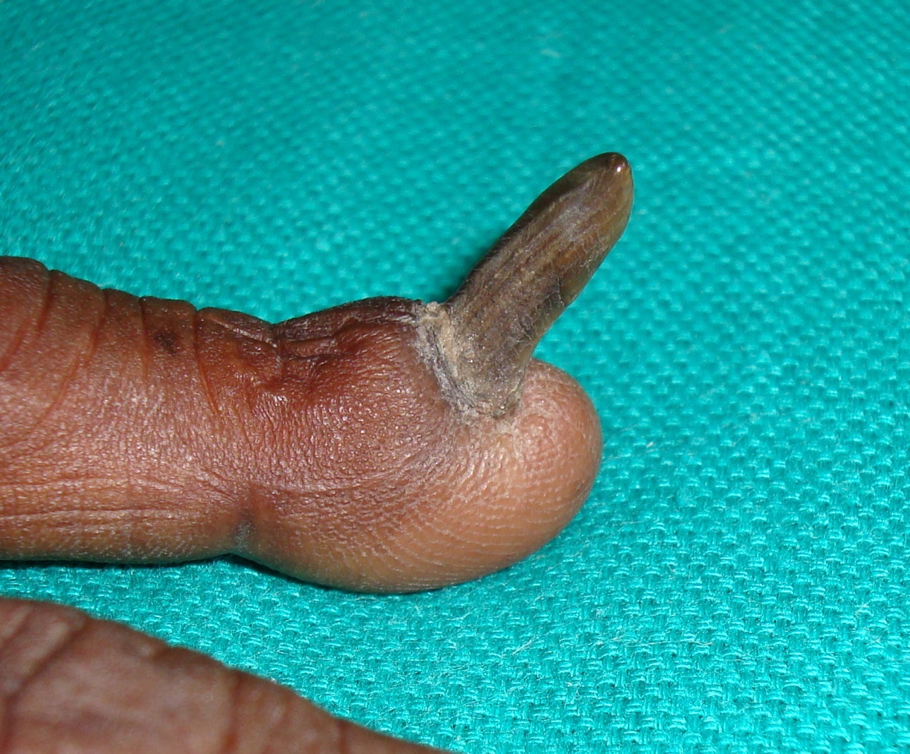
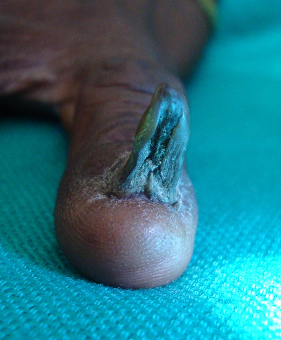
| Figure 1. Onychogryphosis of the fifth fingernail Figure 2. Onychogryphosis of the little fingernail: ventral view | |
On examination, the nail of the left fifth finger was markedly thickened and hyperkeratotic. The cuticle was thick and the proximal nail fold was bulbous. The nail was situated at an angle of approximately 45° to the long axis of the distal phalanx (Figure 1). The nail bed was small and partially deformed with prominent subungual hyperkeratosis. The ventral surface of the nail plate showed a pincer nail appearance around a poorly formed central core (Figure 2). A diagnosis of congenital onychogryphosis of the fifth finger of idiopathic nature was considered. Removal of the entire nail plate followed by chemical destruction of the nail matrix was suggested as treatment to the patient, but he rejected this recommendation.
Discussion
We encountered several diagnostic dilemmas related to the isolated nature of the nail abnormality in our case. There is an absence of any similar descriptions in the literature and our case does not conform to known entities in which this nail abnormality is accompanied by other dermatological or systemic features. We considered following differential diagnosis for our case: idiopathic congenital onychogryphosis, pachyonychia congenita, and yellow nail syndrome.
Onychogryphosis refers to nail plate thickening with gross hyperkeratosis and increased curvature of the nail plate, either downward (oyster-like onychogryphosis) or upward (known as ram’s horn dystrophy) [1, 2]. Onychauxis is thickened nail plate in general without the deformity or change in the curvature of the nail plate [1]. Onychogryphosis occurs most commonly in the toenails of elderly people and is associated with the inability to groom the nails properly or with poorly fitting footwear [1]. It can be associated with psoriasis, ichthyosis, onychomycosis, syphilis, pemphigus, impaired peripheral circulation, diseases of the central nervous system, and repeated nail plate trauma related to foot deformities (hallux valgus/hammer toes/overlapping digits), or it may be idiopathic (acquired or hereditary) [1, 2]. Onychogryphosis of fingernails is uncommon [1].
Hard nails or scleronychia of the fingernails are principally observed in ectodermal dysplasia, pachyonychia congenita (PC), and yellow nail syndrome [3]. The nails become yellowish-brown, usually within months after birth, and show subungual hyperkeratosis with elevation of the nail plate. The nails become progressively thicker and wedge-shaped. In yellow nail syndrome, nails are thickened with increased transverse and longitudinal curvature and a very slow growth rate [3]. The term “hypertrophy of the nail plate” is used to describe nail enlargement and thickening related to effects on the nail matrix (excluding nail bed and hyponychium) [2].
pachyonychia congenita (PC) is an autosomal dominant disorder characterized by onychogryphosis, hyperkeratosis of the palms, soles, knees and elbows, and leukoplakia of the oral mucous membranes [4]. It is caused by mutations in one of four keratin genes, KRT6A, KRT6B, KRT16 or KRT17. Historically, PC was subdivided into two subtypes, PC-1 (Jadassohn-Lewandowski type) and PC-2 (Jackson-Lawler type) on the basis of the clinical presentation alone [5]. But with the identification of the defective genes, it became clear that there is considerable phenotypic overlap between these historical subtypes. Hence, a new molecular genetic classification has been adopted whereby the subtypes of PC refer to the mutated keratin gene. PC-6a then refers to a patient carrying a K6a mutation. PC-6b, PC-16, and PC-17, and named in a similar way. “PC-U” is the designation for cases in which the causative gene is unknown. Clinically, the most striking feature of PC is the painful and debilitating plantar keratoderma. Nail dystrophy presents in variable forms, from very minor or almost absent nail changes to the classic hypertrophic nail dystrophy [5]. In our case, apart from onychogryphosis of a single fingernail, no other features were present to entertain a diagnosis of PC.
The yellow nail syndrome consists of the triad of deformed yellow nails, lymphedema and pleural effusion (no lymphedema or pleural effusion was present in our case) [6].
Congenital onychodystrophy of the index fingers (COIF, Iso-Kikochi syndrome) identifies a clinical syndrome consisting of various nail dysplasias of the index fingers associated with underlying bone abnormalities present at birth [7]. It may be hereditary (autosomal dominant) or sporadic. Nail abnormalities like micronychia, polyonychia, anonychia, hemionychogryphosis and mal-alignment are common in unilateral or bilateral index fingernails [8]. There are deformities of the nails on other fingers and radiographic abnormalities of the distal bony phalanx on the affected fingers. The radiological changes are Y-shaped bifurcation of the distal phalanx on lateral view, narrowing and terminal enlargement of the distal phalanx on anteroposterior view, brachymesophalangia (shortening of the middle phalanx of the fifth digit), and syndactyly of the index and other fingers [7, 8, 9]. Our case did not have features of COIF.
We consider the term “congenital onychogryphosis of the fifth finger” of idiopathic nature to best describes our case. Vertical nail plate growth could be attributed to mal-aligned nail matrix. Visual resemblance of the nail to the leaning tower of Pisa encouraged us to describe it as congenital “leaning tower” nail. Treatment would be surgical removal of the onychogryphotic nail followed by permanent destruction of the nail matrix.
References
1. Rich P, Scher RK. An Atlas of Diseases of the Nail, 1st Edn., New York: The Parthenon Publishing Group Inc 2003: 20.2. Baran R, Dawber RPR, Haneke E, Tosti A. Nail plate and soft tissue abnormalities. Baran R, Dawber RPR, Haneke E, Tosti A, Bristow I, eds. A Text Atlas of Nail Disorders: Techniques in Investigation and Diagnosis, 3rd Edn., London: Martin Dunitz 2003: 100-113.
3. Baran R, Dawber RPR, Haneke E, Tosti A. Nail consistency. Baran R, Dawber RPR, Haneke E, Tosti A, Bristow I, eds. A Text Atlas of Nail Disorders: Techniques in Investigation and Diagnosis, 3rd Edn., London: Martin Dunitz 2003: 166-173.
4. de Berker DAR, Baran R. Disorders of Nails. In: Burns T, Breathnach S, Cox N, Griffiths C, eds. Rook’s Textbook of Dermatology, 8th edn., Oxford: Wiley-Blackwell 2010: 65.20.
5. Wilson NJ, Leachman SA, Hansen CD, McMullan AC, Milstone LM, Schwartz ME, McLean WH, Hull PR, Smith FJ. A large mutational study in pachyonychia congenita. J Invest Dermatol 2011;131:1018-24. [PubMed]
6. Dixit R, Dixit K, Jindal S. Yellow nail syndrome. Lung India 2007;24:66-8.
7. Koizumi H, Tomoyori T, Ohkawara A. Congenital onychodysplasia of the index fingers with anomaly of the great toe. Acta Derm Venereol 1998;78:478-9. [PubMed]
8. Hussein TP, Brandt HR, Gabbi TV, Nico MM. Malformations of the index nails. Clin Exp Dermatol 2009;34:890-1. [PubMed]
9. Kameyoshi Y, Iwasaki Y, Hide M, Yamamoto S. Congenital onychodysplasia of the index fingers in identical twins. Br J Dermatol 1998;139:1120-2. [PubMed]
© 2011 Dermatology Online Journal