Two distinct viral infections complicating pemphigus foliaceus
Published Web Location
https://doi.org/10.5070/D371j1j817Main Content
Two distinct viral infections complicating pemphigus foliaceus
Sarah N Gee MD1, Nicole F Velez MD1, Alireza Sepehr MD2, Susan Burgin MD1
Dermatology Online Journal 18 (1): 3
1. Department of Dermatology2. Department of Pathology
Beth Israel Deaconess Medical Center, Boston, Massachusetts
Abstract
We describe a patient with pemphigus foliaceus who developed two distinct disseminated cutaneous viral infections. Our patient is an 83-year-old female with a recent diagnosis of pemphigus foliaceus, who presented with painful ulcerations while on corticosteroids. Histopathology examination revealed disseminated herpes simplex virus (HSV). Despite adequate treatment with anti-herpetic treatment, some ulcerations failed to heal. A second biopsy revealed the presence of cytomegalovirus (CMV). This was treated successfully with appropriate antiviral therapy. In patients with autoimmune bullous disease, the development of new skin pain or new constitutional symptoms, change in primary morphology, rapid disease progression, or failure to respond to appropriate therapies should prompt the clinician to consider a concurrent cutaneous viral infection. There should be a low threshold to perform ancillary tests, to re-biopsy, and in severe cases, to consider empiric treatment with antiviral treatment therapy and modification of immunosuppressive regimens.
 |  |
| Figure 1 | Figure 2 |
|---|---|
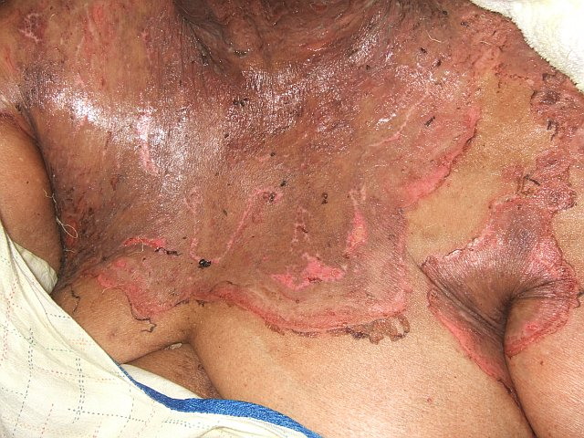
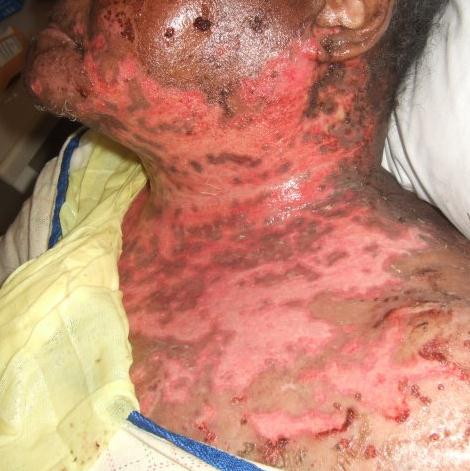
| Figure 1. Anterior chest and neck, at presentation to the emergency department. Figure 2. Twenty-one days after initial presentation showing painful, coalescing, geometric, and friable erosions. | |
An 83-year-old female with type II diabetes and obesity presented to the emergency department with a widespread, pruritic eruption. Examination revealed erythematous, exfoliative plaques with adherent scale on her face, trunk, and extremities (Figure 1). A diagnosis of pemphigus foliaceus (PF) was made based on histopathology and immunofluorescence. Prednisone 60 mg (0.75 mg/kg) daily was initiated.
Seven days after discharge she returned to clinic reporting skin pain. Examination was notable for discrete circular erosions coalescing into large, ulcerative plaques with hemorrhagic crusting (Figure 2). A biopsy from an ulcer edge showed PF with superimposed viral cytopathic changes suggestive of herpes simplex virus (HSV) infection (Figure 3). This diagnosis was confirmed by positive immunostaining and viral culture that grew HSV-Type 2. The patient completed a 2-week course of intravenous acyclovir while continuing prednisone. Re-epithelialization occurred on the head and neck, but lesions persisted on her pannus.
 |  |
| Figure 3 | Figure 4 |
|---|---|
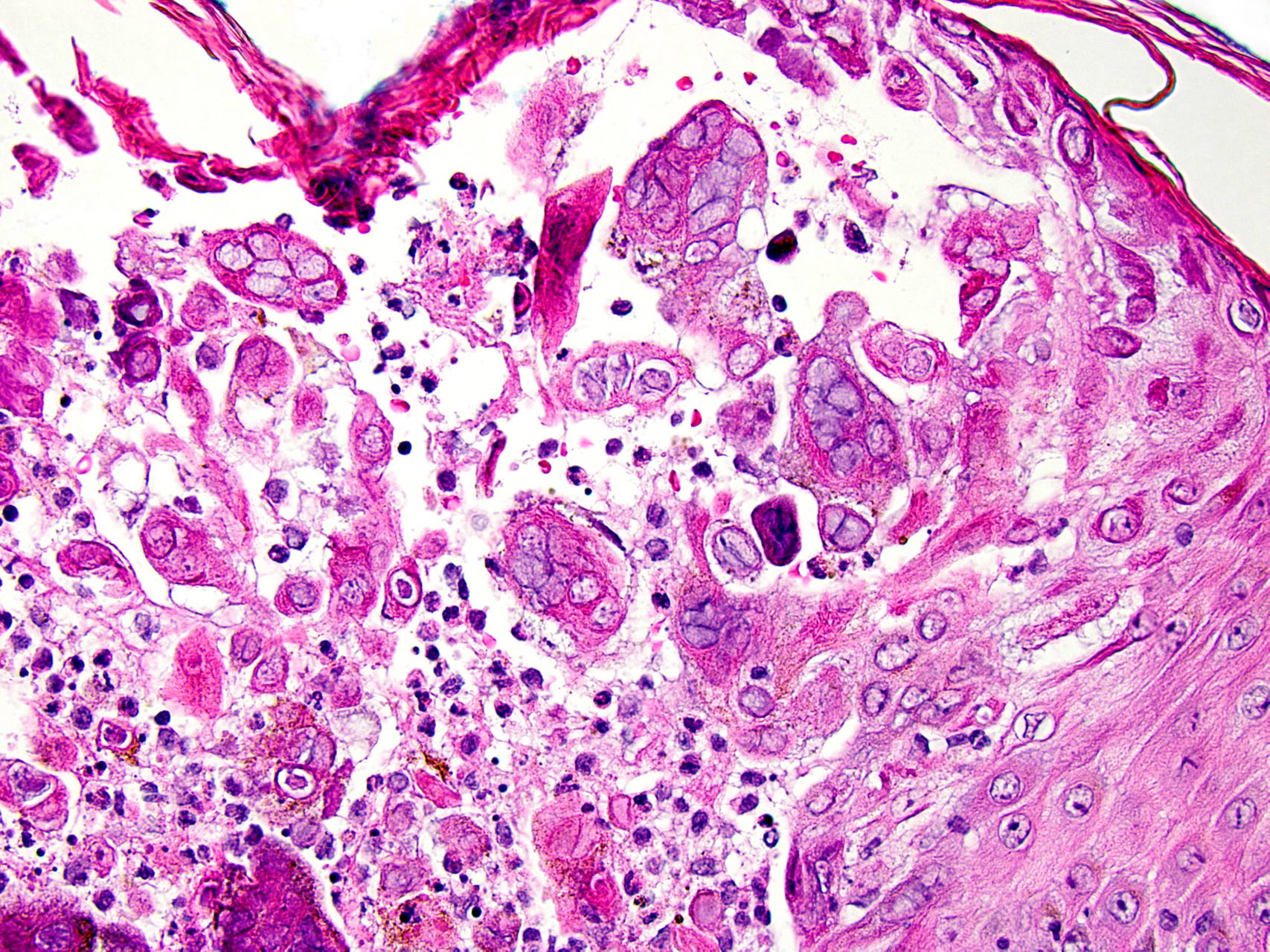
| Figure 3. Hematoxylin and eosin (H&E) stain on the biopsy from right medial upper thigh show marked ballooning degeneration
of keratinocytes and cytopathic changes including “molding,” giant cell formation, and Cowdry bodies consistent with HSV infection
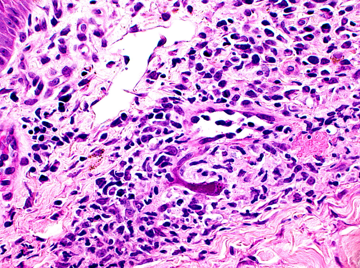
(x200). Figure 4. Superficial, perivascular lympho-plasmacytic infiltrate with vasculopathy and marked viral cytopathic effects of endothelial cells including nuclear and cytoplasmic inclusions suggestive of CMV infection (H&E, x400). | |
Another biopsy was obtained from the pannus. Histopathology revealed viral cytopathic effects of endothelial cells consistent with cytomegalovirus (CMV) (Figure 4). A focal subcorneal blister with mild acantholysis was also noted, suggesting persistent PF activity. The patient's CMV viral load was mildly elevated at 1,590 copies/ml (normal: undetectable). Human immunodeficiency virus antibody was negative. Oral valganciclovir 450 mg twice daily was initiated and prednisone was increased to 80 mg per day (1 mg/kg) for better control of PF. The patient's ulcers healed in two weeks. Repeat CMV viral load was undetectable and she was discharged on prophylactic doses of valganciclovir. Unfortunately, her course was later complicated by multi-bacterial sepsis and she died shortly thereafter.
Our patient is of particular interest because disseminated CMV disease occurred shortly after HSV-induced Kaposi varicelliform eruption (KVE). Whereas KVE is most commonly described in patients with atopic dermatitis, it has also been reported in patients with autoimmune bullous disease. There are 17 reported cases of KVE in PF. Two of these patients died from disseminated HSV viremia, hepatitis, and/or multiorgan failure [1].
Cytomegalovirus cutaneous findings are generally non-specific and can include a morbilliform eruption, purpura, vesicles, verrucous lesions, and ulcerations [2]. In the autoimmune bullous disease population, CMV reactivation is often associated with escalation of immunosuppressive therapy and it can be a significant source of morbidity and mortality. Fever, malaise, and extracutaneous involvement (namely, gastrointestinal) are the most common findings [3, 4, 5]. Casals et al report a case of a patient with bullous pemphigoid who after just two weeks of prednisone therapy developed prolonged fever, which was determined to be secondary to disseminated CMV disease [5]. In one series analyzing the outcomes of pregnant women with pemphigus on high dose prednisone and azathioprine, one intrauterine death occurred because of CMV pneumonitis [6]. In the ophthalmologic literature, CMV retinitis is almost always associated with human immunodeficiency virus (HIV), but reports of otherwise immunocompetent hosts developing CMV retinitis while on systemic prednisone or after treatment with intravitreous corticosteroids do exist [7, 8]. Whereas much has been written in the ophthalmology literature about the risks of ocular CMV disease upon exposure to corticosteroids, we did not find any previous reports of CMV causing cutaneous disease in patients with blistering disease on immunosuppressive therapy.
Because disseminated viral infections can lead to multiple organ involvement and life-threatening disease, it is important to consider the diagnosis in patients with autoimmune bullous diseases with atypical findings or constitutional symptoms. Patients with blistering disease who exhibit (1) changing or atypical skin and/or mucous membranes lesions (i.e., geometric erosions, deeper ulcerations), (2) new skin pain, (3) systemic symptoms (fever, malaise, lymphadenopathy, odynophagia) and/or (4) a lack of response to therapy should be evaluated for cutaneous viral infection [1].
References
1. Demitsu T, Kakurai M, Azuma R, Hiratsuka Y, Yamada T, Yoneda K. Recalcitrant pemphigus foliaceus with Kaposi's varicelliform eruption: report of a fatal case. Clin Exp Dermatol 2008;33:681-2. [PubMed]2. Horn TD, Hood AF. Clinically occult cytomegalovirus present in skin biopsy specimens in immunosuppressed hosts. J Am Acad Dermatol 1989;21:781-4. [PubMed]
3. Choi YL, Kim JA, Jang KT, Kim DS, Kim WS, Lee JH, et al. Characteristics of cutaneous cytomegalovirus infection in non-acquired immune deficiency syndrome, immunocompromised patients. Br J Dermatol 2006;155:977-82. [PubMed]
4. Orton DI, Orteu CH, Rustin MH. Cytomegalovirus-associated gastric ulcer in an immunosuppressed patient with pemphigus vulgaris. Clin Exp Dermatol 2001;26:170-2. [PubMed]
5. Casals DS, Nunes EA, Maruta CW, Aoki V, Santi CG, Simonsen Nico MM, Correa MC, Leite OH, Rivitti EA. Disseminated cytomegalovirus disease as a cause of prolonged fever in a bullous pemphigoid patient under systemic steroid therapy. J Dermatol 2003;30(4):332-6. [PubMed]
6. Kardos M, Levine D, Gurcan HM, Ahmed RA. Pemphigus vulgaris in pregnancy: analysis of current data on the management and outcomes. Obstet Gynecol Surv 2009;64:739-49. [PubMed]
7. Sloan DJ, Taegtmeyer M, Pearce IA, Hart IJ, Miller AR, Beeching NJ. Cytomegalovirus retinitis in the absence of HIV or immunosuppression. Eur J Ophthalmol 2008;18:813-5. [PubMed]
8. Tugal-Tutkun I, Araz B, Cagatay A. CMV retinitis after intravitreal triamcinolone acetonide injection in a patient with Behcet's uveitis. Int Ophthalmol 2010;30:591-3. [PubMed]
© 2012 Dermatology Online Journal