Psoriasis confined to the nails
Published Web Location
https://doi.org/10.5070/D36r0173b5Main Content
Psoriasis confined to the nails
Chrysalyne A Schmults MD
Dermatology Online Journal 9(4): 7
From the Ronald O. Perlman Department of Dermatology, New York University
Abstract
A case is presented of a 49-year-old man with psoriasis limited to the nails for 12 years. The patient had no evidence of psoriasis elsewhere. He had been treated with a year-long course of griseofulvin and 2 months of weekly fluconazole without effect before the diagnosis of psoriasis was made by biopsy of the lateral nail fold. Options for the treatment of nail psoriasis are reviewed.
Clinical summary
History.—A 49-year-old man presented with a 12-year history of nail dystrophy. The patient first noted thickening of his fingernails while residing in his native New Guinea. He was working as a security guard and denied exposure to chemical agents or trauma to his hands. He was treated with multiple topical creams and with oral griseofulvin for 1 year without improvement. He immigrated to the United States in June 2001. A KOH preparation at the initial visit was reported to be positive, and he was treated with 150-200 mg of fluconazole weekly for 2 months without improvement. A nail clipping examined with a periodic-acid Schiff stain was negative for fungi. A fungal culture of nail clippings was negative. A biopsy specimen of the laternal nail fold was obtained. He is currently being treated with clobetasol ointment with mild improvement. His past medical history is unremarkable. He denies pain and pruritus.
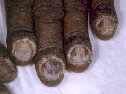
Physical examination.—He had subungual hyperkeratosis of all fingernails and the right first and second toenails. Ridges, crumbling, and onycholysis were also noted. There was hypertrophy of the soft tissues of the distal phalanges and retraction of the cuticles. Clubbing was absent.
|
|
| Figure 1 | Figure 2 |
|---|
Laboratory data.—A complete blood count with differential analysis, liver function tests, electrolyte panel, blood urea nitrogen, creatinine, lipid panel, and stool examination for ova and parasites were normal. A chest radiograph and radiographs of the hands were normal.
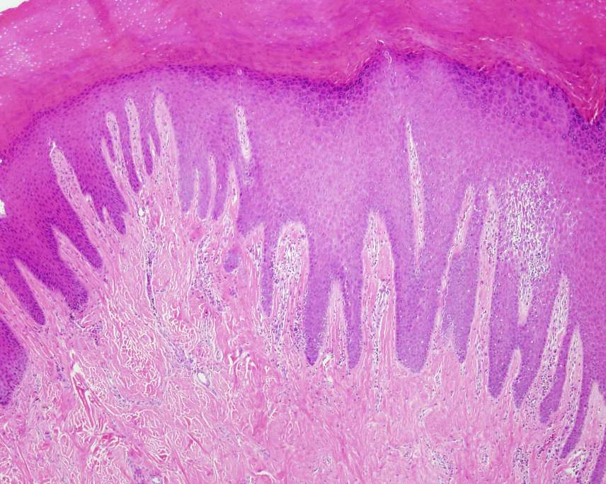
Histopathology.—There is psoriasiform epidermal hyperplasia with thinning of the suprapapillary plates and dilatation of the papillary dermal blood vessels. There are foci of parakeratosis with associated hypogranulosis. A periodic-acid Schiff stain for fungal organisms is negative.
Diagnosis.— Psoriasis confined to the nails.
Comment
Psoriasis limited to the nails is reported to comprise 1-5 percent of patients with psoriasis [1] and is thus an uncommon presentation of a common disease. This patient's case is particularly striking in the severity of the nail involvement.
Psoriasis involving the nail bed gives rise to onycholysis with oil-drop discoloration and subungual hyperkeratosis. Psoriasis of the nail matrix leads to ridges and pits of the nail. Finally, nail-fold psoriasis causes nail-plate crumbling.[2]
Psoriasis of the nails is difficult to treat. Although many dermatologists favor injection of triamcinolone acetonide into the nail matrix via the proximal nail fold and into the nail bed via the lateral nail folds, there are little data to support this treatment. Four studies of intralesional glucocorticoid injection reported improvement ranging from 20-50 percent. However, one study reported 100 percent improvement in hyperkeratosis with 50 percent improvement in pits and onycholysis. [3]
Topical glucocorticoids are commonly used to treat psoriasis of the nail bed but have little effect unless high-potency glucocorticoids are used. Prolonged topical glucocorticoid therapy has led to atrophy of the fingertips. In severe cases, the fingers become tapered with bone loss resulting in a disappearing digit. Other topical therapies include calcipotriol [4], 1 percent 5-fluorouracil [5], 10 percent cyclosporin ointment, anthralin, and tazarotene. These agents are sometimes used in combination with urea or salicylic acid preparations. None of these treatments achieve complete clearance of nail psoriasis, and none is clearly superior. Oral therapies such as PUVA photochemotherapy, cyclosporin, acitretin, and methotrexate have been investigated although these are usually reserved for patients with simultaneous lesions or those with severe pustular psoriasis of the nails.
References
1. Van Laborde S, Scher RK. Developments in the treatment of nail psoriasis, melonychia striata, and onychomyocosis. Dermatol Clin 2000;18:37.2. De Berker D. Management of nail psoriasis. Clin Exp Dermatol 2000;25:357.
3. De Berker D, Lawrence CM. A simplified protocol of nail steroid injection for psoriatic nail dystrophy. Br J Dermatol 1998;138:90.
4. Tosti A, et al. Calcipotriol ointment in nail psoriasis: a controlled double-blind comparison with betamentasome dipropionate and salicylic acid. Br J Dermatol 1998;139:655.
5. De Jong EM, et al. Dystrophic psoriatic fingernails treated with 1 percent 5-fluorouracil in a nail penetration-enhancing vehicle: a double-blind study. Dermatology 1999;199:313.
© 2003 Dermatology Online Journal