Grouped skin metastases from laryngeal squamous cell carcinoma and overview of similar cases
Published Web Location
https://doi.org/10.5070/D364j6g9kxMain Content
Grouped skin metastases from laryngeal squamous cell carcinoma and overview of similar cases
Sadollah Shamsadini MD1, Aliakbar Taheri MD2, Shahriar Dabiri3, Kerman Farshid Darvish Damavandi1, and Siamak Salahi2
Dermatology Online Journal 9(5): 27
Departments of Dermatology1, Otolaryngology2, and Pathology3, Kerman University of Medical Sciences, Kerman, Iran. shamsadini@yahoo.com
Abstract
Cutaneous metastases from internal malignancies or primary skin cancers are uncommon, particularly in a grouped pattern. We report a 58-year-old man with a known case of laryngeal squamous cell carcinoma who underwent radiotherapy after surgical excision of the tumor. Unilateral, grouped, red-brown, vesicle-like nodules appeared on his shoulder 9 months after the laryngeal surgery. The pathologic diagnosis of an excised nodule was metastatic squamous cell carcinoma.
Introduction
In a review of 4,020 patients with metastatic squamous-cell carcinoma (SCC), cutaneous lesions occurred in 10.4 percent [1]. The morphology of these metastases include nodules, ulcers, inflammatory areas, sclerotic areas, bullae, and vesicles [2]. Carcinoma encuirasse, metastases presenting as alopecia, and grouped or zosteriform metastases are additional and uncommon presentations. A literature review found eight cases of grouped zosteriform skin metastases from various primary sites, but none from the larynx [3, 4, 5, 6, 7]. In general, skin metastases of SCC to the head and neck are rare [8]. In a recent description of 2,491 patients with SCC of the upper aerodigestive tract, only 19 (0.763 %) developed skin metastases [8]. As expected, this was a poor prognostic sign; 90 percent of these patients died within a median time of 3 months. We describe a patient with laryngeal SCC who developed grouped vesicle-like metastases on the left shoulder following radiotherapy.

|
|
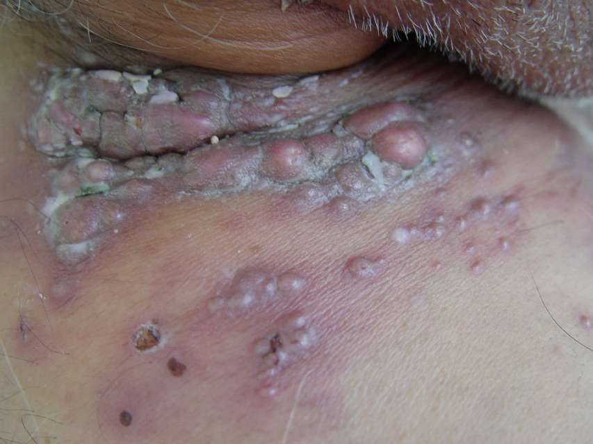
| Figure 1 | Figure 1a |
|---|---|
| Grouped erythematous and vesicle-like nodules | |
|
|
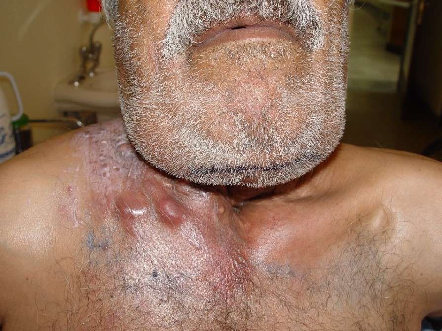
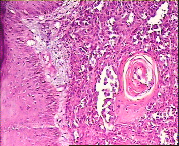
| Figure 1b | Figure 2 |
|---|---|
| Grouped nodules on left shoulder(fig. 1b). Low-power histologic view of metastatic SCC (fig. 2). | |
|
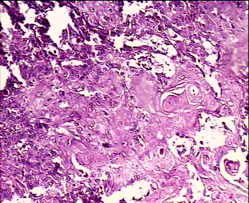
| Figure 3 |
|---|
| Metastatic squamous cell carcinoma. |
Clinical summary
A 58-year-old man with squamous cell carcinoma of the larynx is presented. He had a 20-year history of cigarette smoking and a history of heroin and opium addiction. Laryngeal SCC was diagnosed, and the patient underwent total laryngectomy followed by radiotherapy. Shortly after the radiotherapy and 1 year following surgery, a group of red-violet vesicle-like nodules appeared on his left shoulder. An attempt to aspirate fluid from a shiny, translucent lesion disclosed that it was actually a nodule (figs. 1, 2). A representative lesion on the right shoulder was excised, and the histology revealed a solid tumor with infiltration into the mid-dermis, islands of atypical squamous cells, and a few squamous eddies (fig. 3); these findings were consistent with the diagnosis of a nodular SCC metastasis. Additional radiotherapy was recommended.
Discussion
The clinical appearance of cutaneous metastases varies over a wide morphologic spectrum. Malignant melanoma has a high frequency of metastasis to the skin; skin metastasis from lung and kidney malignancies are less common [9]. In 1998 Cuq-Viguier et al. reported a case with a similar configuration of grouped vesicle-like skin metastases originating from SCC of the stump of an amputated arm [4]. Kato et al. in 2001 reported a case of zosteriform, epidermotropic metastases from primary skin SCC [3]. The cause of a grouped or dermatomal metastatic distribution in malignant lesions is not known, but perineural lymphatic invasion and spread has been hypothesized as a possible explanation for this pattern [10, 11, 12]. Tumor invasion of dorsal root ganglia with peripheral extension may have an important role [13]. Grouped and zosteriform metastases are uncommon but may result from neoplasms of many primary sites. In addition, the upper aerodigestive tract is a rare primary source for cutaneous SCC metastases. Our case demonstrates the unusual combination of grouped cutaneous metastases from a primary laryngeal SCC.
References
1. Lookingbill DP, Spangler N, Helm KF: Cutaneous metastases in patients with metastatic carcinoma: a retrospective study of 4020 patients. J Am Acad Dermatol 1993;29:228-36.2. Brownstein MH, Helwig EB: Spread of tumors to the skin. Arch Dermatol 107:80-6,1973.
3. Kato N,Aoyagi S,Sugawara H,Mayuzumi M.Zosteriform and epidermotropic primary cutaneous squamous cell carcinoma.Am J Dermatopathol Jun 23[3]:216-20 2001
4. Cuq-ViguierL, Viraben R. Zosteriform matastases from squamous cell carcinoma of the stump of an amputated arm. Clin Exp Dermatol. 1998 May;23[3]:116-8.
5. Bauza A, Redonod p,ldoate MA. Cutaneous zosteriform squamous cell carcinoma metastasis arising in an immunocompetent patient. Clin Exp Dermatol. 2002 May;27[3]:199-201.
6. Shafqat A, Viehman GE, Myers SA. Cutaneous squamous cell carcinoma with zosteriform metastasis in a transplant recipient. J Am Acad Dermatol. 1997 Dec;37[6]:1008-9.
7. Manteaux A, Cohen PR, Rapini RP. Zosteriform and epidermotropic metastasis. Report of two cases. J Dermatol Surg Oncol 1992;18:97-100.
8. Pitman KT, Johnson JT. Skin metastases from head and neck squamous cell carcinoma: incidence and impact. Head Neck 1999;21:560-5.
9. Matarasso SL, Rosen T: Zosteriform metastasis:case presentation and review of the literature. J Dermatol Surg Oncol 14: 77-8, 1988.
10. Bluefarb SM, Wallk S, Gecth M: Carcinoma of the prostate with zosteriform cutaneous lesions. Arch Dermatol 76:402-6,1957.
11. Hodge SJ, Mackle S, Owen LG: Zosteriform inflammatory metastatic carinoma. Int Dermatol 18: 142-5, 1979.
12. Shamsadini S Dabiri S Zosteriform metastases in a man with malignant melanoma: Medical journal of the Islamic republic of Iran vol 16 Num 2 August 2002 pp115-17.
13. Jaworsky C, Bergfeld WF: Metastatic transition cell carcinoma mimicking zoster sine herpete. Arch Dermatol 122: 1357-8,1986.
© 2003 Dermatology Online Journal