Granuloma annulare, patch type
Published Web Location
https://doi.org/10.5070/D35z4869nkMain Content
Granuloma annulare, patch type
Frank C Victor MD, Stephanie Mengden MD
Dermatology Online Journal 14 (5): 21
Department of Dermatology, New York UniversityAbstract
A 64-year-old man was referred to the Bellevue Hospital Center Dermatology Clinic for evaluation of an asymptomatic eruption on his left inner arm, which had been present for 4 months and was unresponsive to topical anti-fungal therapy. One month after the initial eruption, 2 similar, asymptomatic lesions appeared on the right inner arm. The lesions were slowly expanding. The histopathology observed in a biopsy specimen from the left medial arm was consistent with interstitial granuloma annulare. The patient's clinical presentation was consistent with patch-type granuloma annulare. He was treated with a mid-potency topical glucocorticoid twice daily for 4 weeks without benefit. Because the eruption was asymptomatic, treatment was discontinued.
Clinical synopsis
A 64-year-old man was referred to the Bellevue Hospital Center Dermatology Clinic for evaluation of an asymptomatic, slowly expanding eruption on his left arm, which had been present for four months. He had been treated with ciclopirox cream twice daily for 3 weeks without improvement. Two months later, a similar asymptomatic eruption was noted on his right arm that was also expanding in size. Past medical history included arthritis for which he took naproxen. He denied other medical problems and he was taking no other medications. He had no known drug allergies. A punch biopsy of the eruption on the left medial arm was performed. The patient was treated with triamcinolone acetonide 0.1 percent ointment twice daily for 4 weeks. The patient noted no improvement in the eruption. Because the lesions were asymptomatic, topical glucocorticoids were discontinued.
Physical Examination
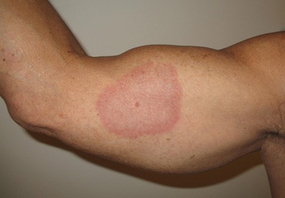
A 9.5 x 9.5 cm, annular, erythematous patch without scale was present on the medial aspect of the left upper arm. Two, non-tender, annular, erythematous patches without scale that measured 2 x 2.5 cm and 2 x 3.5 cm were present on the medial aspect of the right upper arm. The erythema centrally was noted to be less intense than it was at the periphery.
 |  |
| Figure 1 | Figure 2 |
|---|
A complete blood count was normal. A basic metabolic panel was normal except for increased BUN/Cr (31mg/dL/1.5mg/dL). Liver function tests showed a normal aminosuccinyltransferase, an increased aminolysyltransferase level of 40 U/L, and an increased alkaline phosphatase level of 119 U/L. Borrelia burgdorferi antibody was negative.
Histopathology
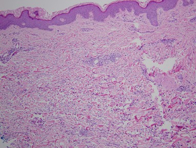
Within the papillary and prominently in the reticular dermis, there is an interstitial infiltrate of histiocytes and lymphocytes with an associated perivascular lymphocytic infiltrate.
Comment
Granuloma annulare (GA) is a cutaneous eruption that occurrs most commonly before the age of 30. It has been noted to occur twice as often in women as compared to men. Several precipitating factors have been proposed, which include sun exposure, insect bites, viral infections, thyroiditis, tuberculin skin tests, and trauma [1, 2, 3]. One hypothesis regarding its pathogenesis is that GA is secondary to a delayed-type hypersensitivity reaction inasmuch as T-helper cells are present in the inflammatory infiltrate. Various cytokines, such as interleukin (IL)-1ß, IL-6, tumor necrosis factor, and metalloproteinases 2 and 9 have been detected [4, 5].
Granuloma annulare most commonly occurs on the extremities, especially the hands and feet although its clinical manifestations are varied [4]. Five morphologic patterns have been described: localized, generalized, perforating, subcutaneous, and patch types [6]. Of these, the localized type is most common and presents as a plaque without scale that is composed of multiple, small papules that coalesce into an arcuate or annular formation. Generalized GA manifests as hundreds to thousands of small, skin-colored-to-erythematous papules that are distributed symmetrically on the extremities and trunk. Some of these papules may coalesce into annular plaques. Papules with central umbilication or crusts are characteristic of perforating GA, which often occurs on the dorsal aspects of the hands and fingers [4]. Subcutaneous GA presents as non-tender, large, subcutaneous nodules with normal overlying skin and is most commonly observed in children [3]. Patch-type GA appears as erythematous patches without scale that may or may not have an annular configuration on the trunk and extremities.
Granuloma annulare is a granulomatous dermatitis histolopathologically with three patterns. The most common is the interstitial pattern, in which histiocytes infiltrate between collagen fibers and minimal degenerated collagen is noted. The second most common is the classic pattern, in which there are palisaded granulomas in the dermis. Degenerated collagen fibers and mucin compose the center of the granulomas and are surrounded by histiocytes and lymphocytes. The last pattern presents as nodules of epithelioid histiocytes and can resemble cutaneous sarcoidosis.
The treatment of GA depends on the presentation as well as on the response to treatment. For localized disease, intralesional glucocorticoids are sometimes effective. Other treatment modalities include topical or oral glucocorticoids, cryotherapy, surgical excision, cyclosporine, fumaric acid esters, chloroquine, and ultraviolet therapy including UVA1 phototherapy [4, 7, 8, 9]. Regarding tumor necrosis factor-alpha inhibitors such infliximab and etanercept, there are reports reflecting resolution of the disease with this treatment modality while others show no benefit [5, 10, 11].
References
1. Mills A, et al. Auricular granuloma annulare: a consequence of trauma? Am J Dermatopathol 1992;14:4312. Muhlbauer JE. Granuloma annulare. J Amer Acad Dermatol 1980;3:217
3. Li A, et al. Granuloma annulare and malignant neoplasms. Am J Dermatopathol 2003;25:113
4. Smith MD, et al. Granuloma annulare. Int J Dermatol 1997;36:326
5. Shupack J, et al. Resolving granuloma annulare with etanercept. Arch Dermatol 2006;142: 394
6. Brey NV, et al. Acute-onset, painful acral granuloma annulare: a report of 4 cases and a discussion of the clinical and histologic spectrum of the disease. Arch Dermatol 2006;142:49
7. Spadino S, et al. Disseminated granuloma annulare: efficacy of cyclosporine therapy. 2006; 19:433
8. Eberlein-Konig B, et al. Disseminated granuloma annulare-treatment with fumaric acid esters. Dermatology 2005;210:223
9. Schnopp C, et al. UVA1 phototherapy for disseminated granuloma annulare. Photodermatol Photoimmunol Photomed 2005;21:68
10. Hertl MS, et al. Rapid improvement of recalcitrant disseminated granuloma annulare upon treatment with the tumour necrosis factor-alpha inhibitor, infliximab. Br J Dermatol 2005;152: 552
11. Kreuter A, et al. Failure of etanercept therapy in disseminated granuloma annulare. Arch Dermatol 2006;142:1236
© 2008 Dermatology Online Journal