Longstanding translucent tumor of the groin
Published Web Location
https://doi.org/10.5070/D35kn8q44wMain Content
Long-standing translucent tumor on the groin.
García-Doval I, De la Torre C, Losada A, Toribio J, Cruces-Prado MJ.
Dermatology Online Journal 5(1): 4
Hospital Provincial de Pontevedra. Spain.
History

|
| Translucent-appearing tumor of the groin |
A 68-year-old man presented with a protruding mass in his groin. He had worked as a blacksmith for fifty years. Thirty-five years ago he had a similar groin lesion biopsied in another hospital. It was diagnosed as "glandular epithelioma". He received soft X-ray therapy. The lesion reappeared five years later, and he was treated with cobalt radiation, with good results. Five years ago a basal cell carcinoma on the back was excised at our hospital. He did not return for follow-up until a slowly growing lesion on his groin, present for the last three years, brought him for consultation. Physical examination revealed a poikilodermatous area on his right groin, with a sessile, translucent-looking, soft tumor, not fixed to deep structures. He also had a lesion on his back diagnosed as Bowen's disease, and a superficial multifocal basal cell carcinoma on his thigh. General physical examination was normal.
|

|
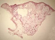
Figure 3: The tumor is composed of "pale-staining lakes, with cellular islands". There are no inflammatory cells. (HE50x)
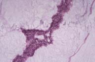
When cut for processing, the groin tumor was described as multinodular, with waxy and violaceus areas. It oozed a mucinous substance.
|
|
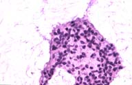
Figure 5: Higher power magnification of the small round cells. Some of the cells show "decapitation" secretion or cytoplasm vacuolation. (HE, 400x)
Laboratory
The following results were negative or normal: Hemogram, biochemistry, chest-X-ray, barium enema, upper gastrointestinal series and thoracoabdominal computerized tomography. Electroneuromyography showed no signs of polyneuropathy. Arsenic levels in nails and pubic hair were normal.
What is your diagnosis?
ANSWER
