Familial reticulate acropigmentation of Dohi: A case report
Published Web Location
https://doi.org/10.5070/D355b0w65vMain Content
Familial reticulate acropigmentation of Dohi: A case report
Maria Princesa P Obieta MD
Dermatology Online Journal 12 (3): 16
Skin and Cancer Foundation, Ortigas Center, Pasig City, Philippines. maprincess@broline.comAbstract
Reticulate acropigmentation of Dohi is a rare dyschromic disorder occurring predominantly among Japanese individuals. Only a few similar cases from Europe and some parts of Asia have been reported. This report describes two Filipino siblings who had reticulate hyperpigmented and hypopigmented macules on the extremities, typical of the disorder.
Introduction
The dyschromatoses comprise a group of autosomal dominant pigmentary disorders characterized by a combination of hyperpigmented and hypopigmented macules forming a reticulate pattern. Two major forms have been described: dyschromatosis universalis hereditaria, the generalized form, and dyschromatosis symmetrica hereditaria (DSH), the localized form. DSH is also called reticulate acropigmentation of Dohi.
Clinical synopsis
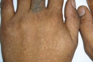
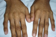
A 26-year-old Filipino man and his 15-year-old sister presented with asymptomatic and progressive mixtures of hyperpigmented and hypopigmented macules affecting the limbs in a reticulate pattern.
 |  |
| Figure 1A | Figure 1B |
|---|---|
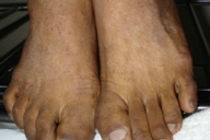
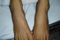
| Figure 1. Reticulate hyperpigmented and hypopigmented macules on dorsa of hands in male (A) and female (B) sibling. (A tattoo is present on one finger in the first patient.) | |
 |  |
| Figure 2A | Figure 2B |
|---|---|
| Figure 2. Similar pigmentation is seen on the dorsa of feet in male (A) and female (B) sibling. | |
 |
| Figure 3 |
|---|
| Family pedigree. |
The lesions were said to have appeared in infancy on the backs of the hands and feet, and slowly progressed to involve the dorsal and ventral surfaces of the extremities. The face and trunk were spared. Many of their relatives had the same lesions with similar distribution.
Examination revealed no palmar punctiform pits or breaks in the epidermal rete ridge patterns. There were no other cutaneous abnormalities noted.
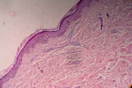
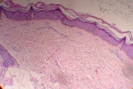
Two skin biopsies were obtained from the older sibling. The first specimen taken from the hypopigmented macule showed decreased melanin pigments at the basal layer. The second specimen taken from the hyperpigmented macule showed increased melanin pigments at the epidermis.
Comment
Reticulate acropigmentation of Dohi (RAD) is characterized by the presence of hyperpigmented and hypopigmented pinpoint or pea-sized macules over the dorsa of the hands and feet and occasionally on the arms and legs [1]. Lesions usually appear in infancy or early childhood [2]. Histology of hyperpigmented lesions shows increased melanin pigments at the basal layer. Decreased or absent melanin pigment is observed in hypopigmented lesions. The condition belongs to a group of autosomal dominant reticulate pigmentary disorders that also includes reticulate acropigmentation of Kitamura (RAPK), and Dowling-Degos disease (DDD). A number of authors suggest that RAPK and DDD are clinical variants of the same entity [3].
RAPK typically presents during the first or second decade of life as a slowly progressive network of hyperpigmented macules on the extensor surfaces of the hands and feet. Punctiform pits and breaks in the epidermal rete ridge pattern are often present. In contrast to the findings in RAD, hypopigmented macules are not observed. The abnormality is characterized histologically by epidermal atrophy and an increased number of basal melanocytes [2, 4].
DDD [2, 4] is a slowly progressive genodermatosis that manifests as reticulate hyperpigmented macules in flexural areas including the axillae, groin, inframammary areas, neck, and rarely the wrists, face, arms, and scalp. The onset is usually in the fourth decade of life. Dark comedo-like lesions on the neck and pitted perioral scars are other features of this disease. Pigmented filiform epidermal projections are seen histopathologically. Basal melanocytes are unaffected in number.
The cause and pathogenesis of RAD have not been defined. Studies indicate various mutations in the RNA-specific adenosine-deaminase gene to be associated with this disorder [5]. These patients are the first cases of reticulate acropigmentation of Dohi reported from the Philippines.
References
1. Urabe K, Hori Y. Dyschromatosis. Semin Cutan Med Surg. 1997;161: 81-85.2. Alfadley A, Al Ajlan A, Hainau B. Reticulate acropigmentation of Dohi: A case report of autosomal recessive inheritance. J Am Acad Dermatol 2000;43:113-117.
3. Dhar S, Kanwar AJ, Jebraili R, Dawn G, Das A. Spectrum of reticulate flexural and acral pigmentary disorders in northern India. J Dermatol. 1994;21:598-603.
4. Danese P, Zanca A, Bertazzoni MG. Familial reticulate acropigmentation of Dohi. J Am Acad Dermatol 1997;37:884-886.
5. Miyamura Y, Suzuki T, Kono M, et al. Mutations of the RNA-specific adenosine deaminase gene (DSRAD) are involved in dyschromatosis symmetrica hereditaria. Am J Hum Genet. 2003;73:693-699.
© 2006 Dermatology Online Journal


