Brown plaques on the lower back
Published Web Location
https://doi.org/10.5070/D35431j6w6Main Content
Unknown: Brown plaques on the lower back
Tiago Esteves MD, Lurdes Ferreira, Isabel Viana, Olívia Bordalo
Dermatology Online Journal 16 (6): 11
Hospital Central do Funchal, Funchal, Madeira, Portugal. tiago.castroesteves@gmail.com
|
|
| Figure 1 | Figure 2 |
|---|---|
|
|
|
|
| Figure 3 |
|---|
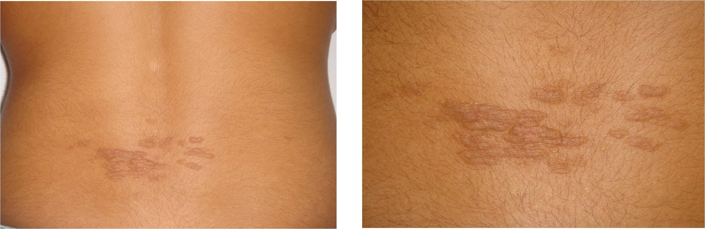
A 12-year-old girl presented with a 2-year history of progressive evolution of multiple, asymptomatic brown plaques on the lower back (Figure 1). These plaques gradually enlarged and increased in number. The patient was otherwise healthy and had received no treatment before coming to our clinic.
On physical examination, we observed multiple brown plaques of coalescent, linearly arranged papules on the lower back. The plaques were soft, compressible and well-defined, with a zosteriform distribution. There was no history of antecedent trauma to the area or family history of similar lesions. No other physical or systemic abnormality was detected. Routine laboratory investigations including thyroid function tests and protein electrophoresis were normal.
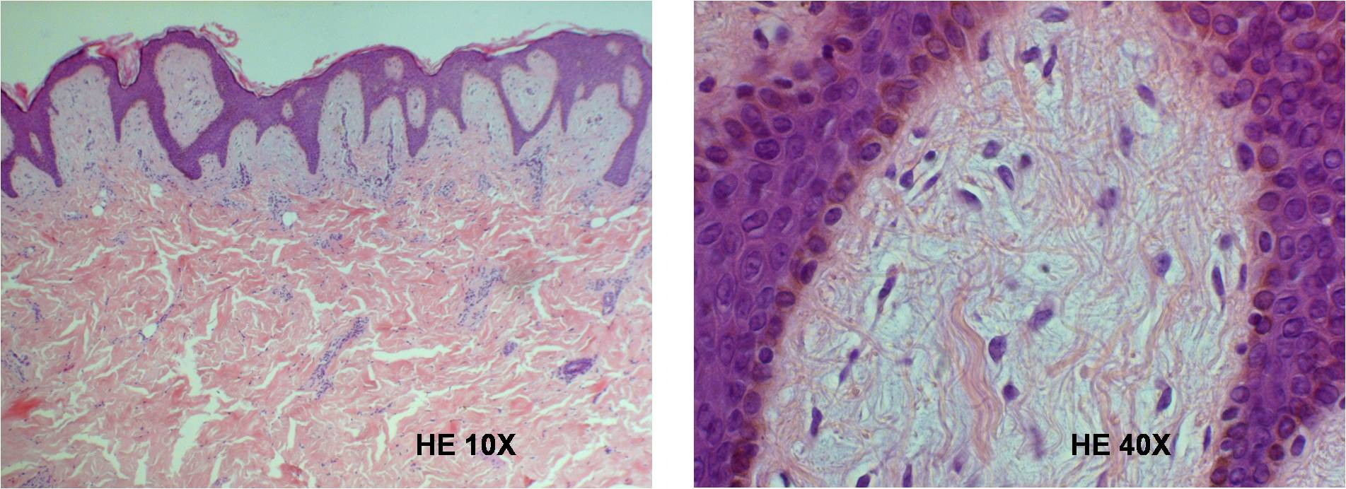
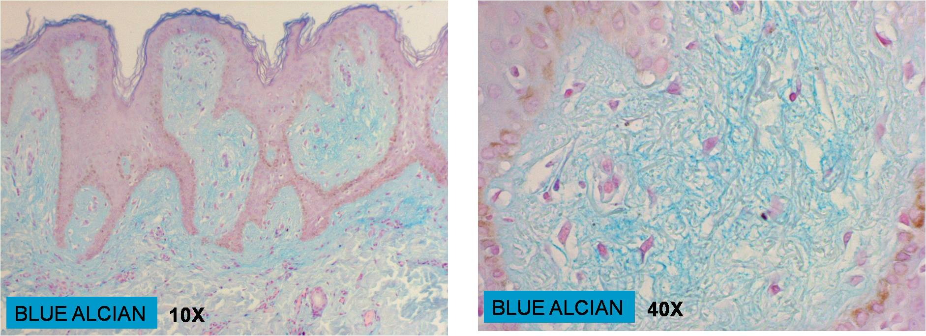
A biopsy of the lesion was performed and the histopathological examination revealed hyperkeratosis, papillomatosis, and elongated rete ridges in the epidermis (Figure 2). On special staining (Alcian blue), mucin deposition was seen in the papillary dermis (Figure 3). The remaining dermis was normal.
What is your diagnosis?
Answer
© 2010 Dermatology Online Journal