Interferon induced sarcoidosis with cutaneous involvement along lines of venous drainage in a former intravenous drug user
Published Web Location
https://doi.org/10.5070/D34wg417hbMain Content
Interferon induced sarcoidosis with cutaneous involvement along lines of venous drainage in a former intravenous drug user
Fareesa Shuja MD, Shaheen C Kavoussi MD, Mohsin R Mir MD, Reena P Jogi MD, Ted Rosen MD
Dermatology Online Journal 15 (12): 4
Department of Dermatology, Baylor College of Medicine, Houston, Texas. vampireted@aol.comIntroduction
Interferons are cytokines with anti-viral and anti-oncogenic properties which, in combination with ribavirin, have become the standard of care for chronic hepatitis C virus (HCV) infection by bolstering the CD4+ T-helper type 1 (Th-1) response [1]. An undesirable consequence of this is the promotion of granuloma formation via the differentiation of mononuclear cells into epithelioid cells, the activation of macrophages, and the upregulation of IL-12 production [2, 3]. Interferon-induced cutaneous granulomatous reactions are rare but have been well-described; systemic sarcoidosis presenting as a cutaneous granulomatous reaction is rarer still but does occur. Here we discuss a case of systemic sarcoidosis presenting as a delayed foreign body granulomatous reaction along lines of venous drainage in a former intravenous drug user. Only one other case of interferon-induced cutaneous sarcoidosis presenting along lines of injection drug use was found in the readily available literature [4].
Case presentation
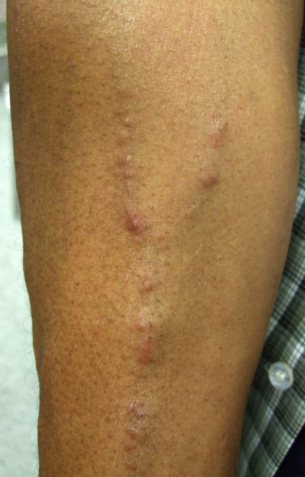 |
| Figure 1 |
|---|
| Figure 1. Right forearm with firm skin-colored to reddish brown nodules along the course of the veins |
A 59-year-old African-American male with HCV-related chronic active hepatitis and a remote history of injection drug use presented with a complaint of nodules developing along the superficial veins of his right forearm for several months. The patient had been receiving treatment with pegylated interferon-α-2b for the past two years. His physical examination was remarkable for scattered, non-tender, firm, reddish-brown nodules along the course of the veins of his right hand and forearm (Fig. 1).
 |  |
| Figure 2 | Figure 3 |
|---|---|
| Figure 2. Skin biopsy specimen; granulomatous infiltrate involving the dermis (H&E, x100) Figure 3. Multiple non-caseating granulomas composed of epithelioid histiocytes and giant cells (H&E, x200) | |
A shave biopsy was performed and histologic examination revealed granulomatous inflammation with polarizable foreign material (Figs. 2 & 3). Angiotensin-converting enzyme (ACE) level was markedly elevated at 153 U/L (reference range 3-48 U/L). A chest radiograph did not reveal any hilar lymphadenopathy, and a urine drug screen was negative.
The patient remained on interferon therapy as his hepatitis C viral load was deceasing in response to treatment. However, several months later when an esophagogastroduodenoscopy (EGD) was performed to screen for varices, biopsies of the antrum and body of the stomach revealed multiple granulomas supporting the diagnosis of systemic sarcoidosis. A gallium scan subsequently showed abnormal uptake in both lungs suggestive of parenchymal pulmonary involvement. The interferon therapy was discontinued, and the patient was referred for additional pulmonary and ophthalmologic evaluation
Discussion
At least 2.7 million Americans are infected with chronic hepatitis C, and an increasing proportion of these patients are being treated with INF-α [5]. The first case of sarcoidosis in a patient receiving interferon was described by Abdi et al. in 1987 [6]. Since then, interferon-induced sarcoidosis has been detailed in numerous cases in the readily available literature [1, 4-18].
Marcoval and his group investigated cutaneous granulomatous lesions in patients with systemic sarcoidosis and found that 22 percent of the patients had foreign material present [19]. Any foreign material can initiate a foreign body immune response; for instance, scars [1, 7], hyaluronic acid injection sites [8], and tattoos [9, 10, 11, 12] have all served as a nidus for granuloma formation in patients receiving INF-α. Fischer et al. describe a patient with cutaneous sarcoidosis ten weeks after treatment with IFN-α-2a and ribavirin and ten years after injection of Artecoll, a permanent soft tissue filler containing insoluble polymethylmethacrylate [13]. The constellation of clinical and histologic findings seen in our patient suggests systemic sarcoidosis with a cutaneous foreign body granulomatous reaction to particulate foreign matter, likely talc, injected some thirty years earlier.
Remissions and reductions of cutaneous lesions have been described as being directly related to discontinuation of interferon, with 35 percent of patients requiring the addition of systemic steroids; unfortunately, the latter can increase the HCV viral load [14]. Dermatologists can play a pivotal role in identifying the early cutaneous manifestations of sarcoidosis. By discontinuing INF-α treatment, whenever feasible, we may be able to prevent the need for steroids at a later time, ultimately averting exacerbation of the initial diagnosis, hepatitis C infection.
References
1. Eberlein-Konig B, Hein R, Abeck D, Engst R, et al. Cutaneous sarcoid foreign body granulomas developing in sites of previous skin injury after systemic interferon-alpha treatment for chronic hepatitis C. Br J Dermatol 1999; 140(2): 370-372. [PubMed]2. Moller DR. Cells and cytokines involved in the pathogenesis of sarcoidosis. Sarcoidosis Vasc Diffuse Lung Dis 1999; 16(1): 24-31. [PubMed]
3. Moller DR, Forman JD, Liu MC, Noble PW, et al. Enhanced expression of IL-12 associated with Th-1 cytokine profiles in active pulmonary sarcoidosis. J Immunol 1996; 156(12): 4952-4960. [PubMed]
4. Doyle MK, Berggren R, Magnus JH. Interferon-induced sarcoidosis. J Clin Rheumatol 2006; 12(5): 241-248. [PubMed]
5. Hurst EA, Mauro T. Sarcoidosis associated with pegylated interferon alfa and ribavirin treatment for chronic hepatitis C: a case report and review of the literature. Arch Dermatol 2005; 141(7): 865-868. [PubMed]
6. Abdi EA, Nguyen GK, Ludwig RN, Dickout WJ. Pulmonary sarcoidosis following interferon therapy for advanced renal cell carcinoma. Cancer 1987; 59(5): 896-900. [PubMed]
7. Perez-Gala S, Delgado-Jimenez Y, Goiriz R, Fernandez-Herrera J, et al. Cutaneous sarcoidosis limited to scars following pegylated interferon alfa and ribivirin therapy in a patient with chronic hepatitis C. J Eur Acad Dermatol Venereol 2007; 21(3): 393-394. [PubMed]
8. Descamps V, Landry J, Frances C, Marinho E, et al. Facial cosmetic filler injections as possible target for systemic sarcoidosis in patients treated with interferon for chronic hepatitis C: two cases. Dermatology 2008; 217(1): 81-84. [PubMed]
9. Nawras A, Alsolaiman MM, Mehboob S, Bartholomew C, et al. Systemic sarcoidosis presenting as a granulomatous tattoo reaction secondary to interferon-alpha treatment for chronic hepatitis C and review of the literature. Dig Dis Sci 2002; 47(7): 1627-1631. [PubMed]
10. Toulemonde A, Quereux G, Dreno B. Sarcoidosis granuloma on a tattoo induced by interferon alpha. Ann Dermatol Venereol 2004;131(1): 49-51. [PubMed]
11. Perera GK, Calonje E. Systemic sarcoidosis presenting in a tattooed man undergoing treatment for hepatitis C. Clin Exp Dermatol 2006; 31(3): 387-389. [PubMed]
12. Werchniak AE, Cheng, SX, Dhar AD, Klaus SN. Sarcoidosis presenting as tattoo changes in a patient undergoing treatment with interferon-alpha and ribivirin. Clin Exp Dermatol 2004; 29(5): 547-548. [PubMed]
13. Fischer J, Metzler G, Schaller M. Cosmetic permanent fillers for soft tissue augmentation: a new contraindication for interferon therapies. Arch Dermatol 2007; 143(4): 507-510. [PubMed]
14. Ramos-Casals M, Mana J, Nardi N, Brito-Zeron P, et al. Sarcoidosis in patients with chronic hepatitis C virus infection: analysis of 68 cases. Medicine (Baltimore) 2005; 84(2): 69-80. [PubMed]
15. Tebben PJ, Atkinson JL, Scheithauer BW, Erickson D. Granulomatous adenohypophysitis after interferon and ribavirin therapy. Endocr Pract 2007; 13(2): 169-175. [PubMed]
16. Yan KK, Dinihan I, Freiman J, Zekry A. Sarcoidosis presenting with granulomatous uveitis induced by pegylated interferon and ribavirin therapy for hepatitis C. Intern Med J 2008; 38(3): 207-210. [PubMed]
17. Adla M, Downey KK, Ahmad J. Hepatic sarcoidosis associated with pegylated interferon alfa therapy for chronic hepatitis C: case report and review of literature. Dig Dis Sci 2008; 53(10): 2810-2812. [PubMed]
18. Cogrel O, Doutre MS, Marliere V, Beylot-Barry M, et al. Cutaneous sarcoidosis during interferon alfa and ribavirin treatment of hepatitis C virus infection: two cases. Br J Dermatol 2002; 146(2): 320-324. [PubMed]
19. Marcoval J, Mañá J, Moreno A, Gallego I, et al. Foreign bodies in granulomatous cutaneous lesions of patients with systemic sarcoidosis. Arch Dermatol 2001; 137(4): 427-30. [PubMed]
© 2009 Dermatology Online Journal