Necrotic Erythema Nodosum Leprosum (NENL)
Published Web Location
https://doi.org/10.5070/D34mm3r7h9Main Content
Necrotic erythema nodosum leprosum
Vandana Mehta Rai MD DNB, C Balachandran MD
Dermatology Online Journal 12 (3): 12
Department of Skin and STD, Kasturba Medical College, Manipal, Karnataka. vandanamht@yahoo.comAbstract
A 32-year-old fisherman suffering from lepromatous leprosy presented with fever, joint pains, bullous lesions, and multiple well-defined ulcers on the extremities and back of 10-days duration. The bullae ruptured to form ulcers of varying sizes with well-defined margins and necrotic bases. A few of the ulcers had black crusts resembling eschars. An immunofluorescence study ruled out autoimmune bullous disease. Histopathology of a lesion showed features of erythema nodosum leprosum.
Introduction
Erythema nodosum leprosum (ENL), also known as type-2 reaction, is characterized by brightly red, raised, tender nodules and plaques of varying sizes. They are warmer than the surrounding skin and they blanch on light touch. The severe from is more common in caucasians and mongolians. Necrotic and ulcerative forms are the rare presentations of severe ENL. Necrotic ENL (NENL) poses diagnostic difficulty in leprosy patients because it mimics other cutaneous necrotizing disorders.
Clinical synopsis
 |  |
| Figure 1 | Figure 2 |
|---|---|
| Figure 1. Close-up view of patient | |
Figure 2. Left arm | |
 |  |
| Figure 3 | Figure 4 |
|---|---|
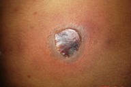
| Figure 3. Bullous lesion on the shoulder | |
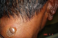
Figure 4. Nodular infiltration of the ear | |
 |  |
| Figure 5 | Figure 6 |
|---|---|
| Figure 5. Bullae on the back and ear | |
Figure 6. Back showing necrosis | |
 |
| Figure 7 |
|---|
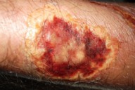
| Figure 7. Hemorrhagic necrosis |
A 32-year-old male fisherman suffering from lepromatous leprosy and on multibacillary multidrug therapy for the past 2 years presented with complaints of multiple ulcers and bullous lesions on the upper and lower extremities for 10 days. The patient had been suffering from recurrent episodes of ENL for the past 6 months for which he was on tapering doses of steroids. The present complaints started with mild fever and joint pains followed by development of multiple erythematous, tender nodules on the extremities and trunk. Within a few days the nodular lesions became bullous; over the subsequent next 2-3 days the bullae ruptured leaving ulcers covered with an eschar-like black hemorrhagic crust. There was no history suggestive of neuritis, orchitis, iritis, arthritis, or glomerulonephritis. Cutaneous examination revealed nodular infiltration of the ear lobes, madarosis, and multiple tense and flaccid bullae on the extremities and trunk. There were multiple ulcers of varying sizes with well-defined margins and necrotic bases. There were multiple atrophic scars of varying shapes and sizes and hyperpigmented macules on the legs and thighs. Nerves including the greater auricular, ulnar, radial, radial cutaneous, ulnar cutaneous, and common peroneal were thickened bilaterally and nontender. Motor examination was normal. The patient had glove-and-stocking anesthesia.
Hematological examination revealed leukocytosis with raised ESR. A slit-skin smear for acid-fast bacilli showed a 4+ bacterial index (BI) and morphological index (MI) of 0 percent. Skin biopsy revealed epidermal spongiosis with neutrophilic infiltration of the dermis and neural and periadnexal infiltration by foamy macrophages, epithelioid cells and neutrophils. Fite stain demonstrated fragmented acid-fast bacilli. Both direct and indirect immunofluorescence studies did not show features of autoimmune bullous disorders. The patient was managed with high-dose steroid and antibiotics; the ENL lesions improved.
Discussion
Reactions in leprosy are divided into two types. Type-1 reaction is associated with alterations in cell-mediated immunity and is seen in tuberculoid and borderline groups; type-2 lepra reaction occurs in lepromatous leprosy. ENL in addition to the other features of systemic involvement such as arthritis, myositis, and nephritis, is the most characteristic lesion of type-2 reaction, although it is not seen in all cases [1]. Classic ENL is characterized by the appearance of crops of tender painful subcutaneous nodules that may be associated with fever and other constitutional symptoms. As the lesions subside they become purplish. Ulceration may occur and lesions heal with scarring. ENL is encountered during therapy of patients with lepromatous leprosy or borderline lepromatous leprosy and may occur as the presenting manifestation of the disease [2.] Vesiculobullous, pustular, ulcerated, hemorrhagic and erythema multiforme-like lesions have been reported in ENL [3]. A bullous-type reaction mimicking pemphigus in lepromatous leprosy has been reported from India however the patient did not show neuritis or ENL lesions, and histopathological examination and immunofluorescence studies were not performed to support the diagnosis [4]. Necrotic ulcerative lesions of long standing untreated Lucio leprosy of Mexicans can be mistaken for necrotic ENL. The lesions of Lucio phenomenon appear on the extremities as painful tender red patches on the skin, which become purpuric; the centers of the lesions develop necrotic ulcers covered with black crust healing with superficial atrophic scars. Excellent results are seen with rifampin and good results with dapsone and steroids. NENL appears in crops along with constitutional symptoms such as fever, joint pain, and malaise, and it occurs in treated patients of lepromatous leprosy. NENL responds very well to thalidomide as well as to systemic steroids but not to rifampin and dapsone. Cutaneous necrotizing vasculitis can also present as similar necrotic lesions. They can be differentiated by the absence of other features of leprosy, negative slit skin smears, and negative findings in tissue biopsy [5.]
References
1. Jopling WH. Reactions in leprosy. Lep Rev 1970;41:62-632. Gomathy Sethuraman, Divakaran Jeevan, Chakravarthy Rangachary Srinivas. Bullous erythema nodosum leprosum (bullous type 2 reaction). Int J of Dermatol 2002;41:363-364
3. Verma KK, Pandhi RK. Necrotic erythema nodosum leprosum; a presenting manifestation of lepromatous leprosy. Int J Lepr other Mycobact Dis 1993;61:293-4.
4. Petro TS. Bullous type of reaction mimicking pemphigus in lepromatous leprosy. Indian J Lepr 1996;68:179-81
5. K Deb Barman, U Gupta, K Saify. Necrotic erythema nodosum leprosum. Indian J Lepr 2005;77:169-172.
© 2006 Dermatology Online Journal

