A case of multiple epithelioid angiomatous nodules
Published Web Location
https://doi.org/10.5070/D34439p27kMain Content
A case of multiple epithelioid angiomatous nodules
Imen Labbène1 MD, Soumaya Rammeh2 MD, Nadia Znaidi2 MD, Becima Fazaa1 MD, Rachida Zermani2 MD
Dermatology Online Journal 18 (8): 8
1. Dermatology Department, Charles Nicolle Hospital Tunis, Tunisia2. Pathology Department, Charles Nicolle Hospital Tunis, Tunisia
Abstract
Cutaneous epithelioid angiomatous nodule (CEAN) is a distinct type of epithelioid vascular tumor that is usually solitary. Herein we present a 31-year-old man with multiple, rapidly growing nodules on the scalp.
Introduction
Cutaneous epithelioid angiomatous nodule (CEAN) is a new and distinct type of epithelioid vascular lesion located in the superficial dermis [1]. According to the original series of 15 cases of CEAN, the clinical presentation was that of a solitary, fast-growing papule or nodule predominantly located on the trunk and extremities.
We present an unusual case of multiple epithelioid angiomatous nodules clustered at the left side of the scalp.
Case report
A 31-year-old man with no relevant history of disease visited our department with multiple rapidly growing nodules that developed over a 2-year period on the scalp.
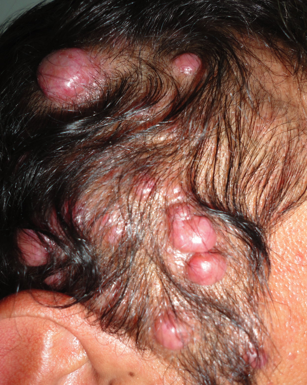
The physical examination revealed 13 erythematous nodules clustered at the left side of the scalp. The lesions varied in size from 5 to 20 mm in diameter (Figure 1).
 |  |
| Figure 1 | Figure 2 |
|---|---|
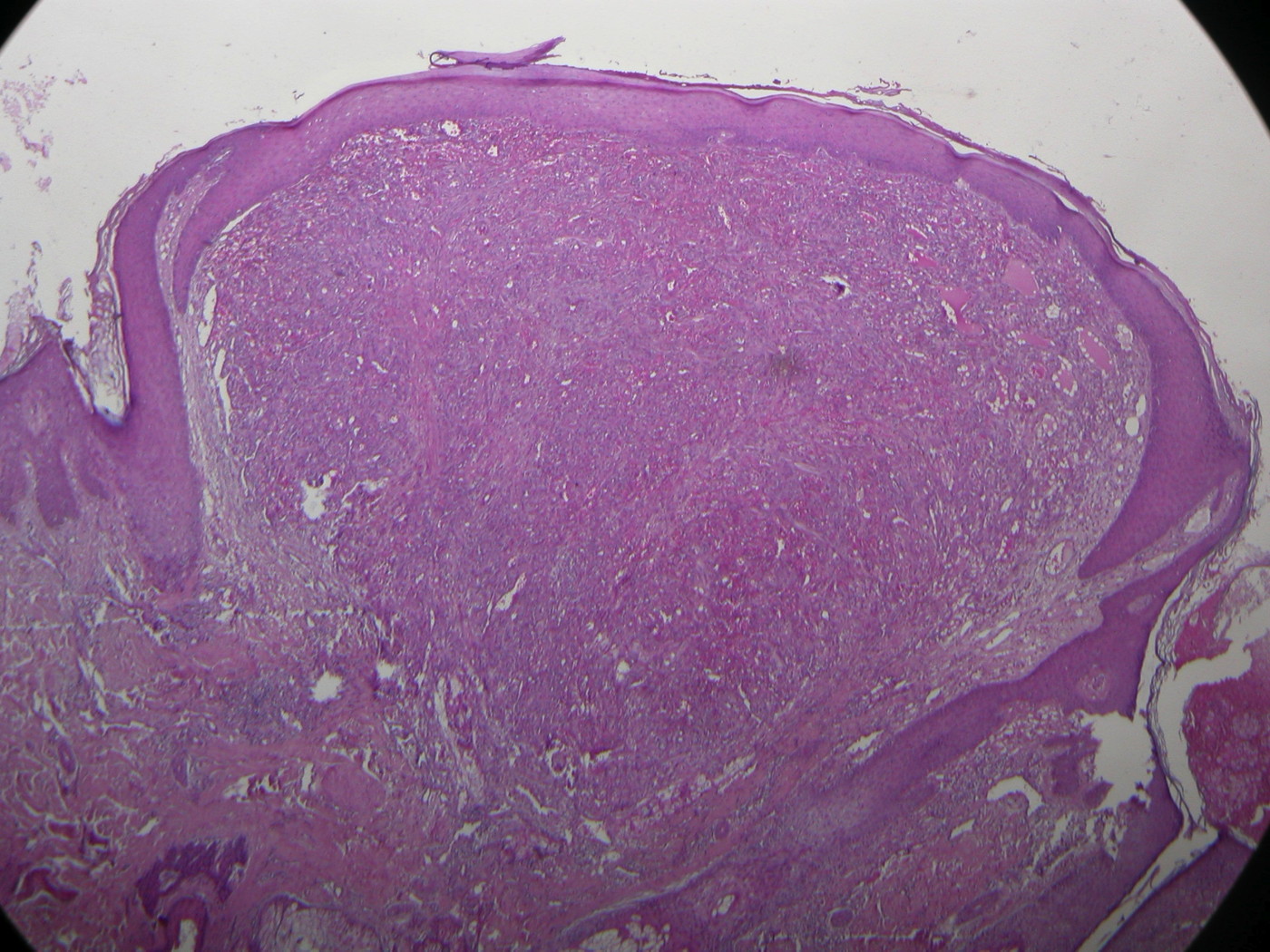
| Figure 1. Multiple erythematous nodules on the scalp Figure 2. Well-circumscribed nodule in the superficial dermis (H&E x40) | |
A nodule was resected and the histology revealed a well-circumscribed unilobular lesion in the superficial dermis (Figure 2) composed of solid sheets of large epithelioid endothelial cells with abundant eosinophilic cytoplasm, enlarged nuclei, and prominent nucleoli (Figure 3). Dispersed within the nodule were open channels (Figure 4) displaying a single cell-lined epithelioid endothelium with some intracytoplasmic vacuoles. There were also a few scattered stromal lymphocytes and eosinophils. No capsule or pseudocapsule was identified. Mitotic figures were present but neither atypical mitoses nor nuclear atypia were seen. The overlying epidermis was unaltered.
 |  |
| Figure 3 | Figure 4 |
|---|---|
| Figure 3. The tumor is composed of epithelioid endothelial cells with abundant eosinophilic cytoplasm. Some endothelial cells
display intracytoplasmic vacuoles (H&E x400). Figure 4. Intralesional vascular channels within the nodule (H&E x100). | |
Immunostaining was positive for Ki-67 in up to 20 percent of the cells and tests for CD31 and CD34 were also positive.
Discussion
CEAN is a benign vascular proliferation that was first described in 2004 by Brenn and Fletcher [1] in a study of 15 cases. It is a rare entity and, to our knowledge, only 51 cases have been reported to date [2].
Clinically, typical lesions usually present as solitary, small reddish papules or nodules of short duration located predominantly on the trunk or limbs of adults. Of the 51 patients reported, 46 had solitary lesions and 5 patients had multiple lesions clustered at the same anatomic site as in our case. In all cases described to date, excision was curative without recurrence.
The growth consists of a solid, often well-circumscribed, proliferation of large epithelioid endothelial cells with frequent intracytoplasmic vacuoles in the superficial dermis. Intralesional endothelial-lined channels are a focal but constant feature. The epithelioid cells are positive for endothelial markers such as CD31, CD34, and von Willebrand factor. Mitotic activity is variable (up to 5/10 HPF). However, atypical mitoses are absent [1, 3].
Although some authors suggested that CEAN should be classified as a solid variant of epithelioid hemangioma [3, 4], others support that CEAN represents a distinct entity in the spectrum of epithelioid vascular proliferations that defies classification according to current criteria [5, 6].
Cutaneous epithelioid angiomatous nodule must be differentiated from other epithelioid vascular proliferations, especially those with malignant behavior such as epithelioid hemangioendothelioma and epithelioid angiosarcoma.
References
1. Brenn T, Fletcher CD. Cutaneous epithelioid angiomatous nodule: a distinct lesion in the morphologic spectrum of epithelioid vascular tumors. Am J Dermatopathol. 2004; 26; 14-21. [PubMed].2. Pavlidakey PG, Burroughs C, Karrs T, Somach SC. Cutaneous epithelioid angiomatous nodule: a case with metachronous lesions. Am J Dermatopathol. 2011 Dec; 33(8):831-4. [PubMed].
3. Sangueza OP, Walsh SN, Sheehan DJ, Orland AF, Llombart B, Requena L. Cutaneous epithelioid angiomatous nodule: a case series and proposed classification. Am J Dermatopathol. 2008; 30;16-20. [PubMed].
4. Weiss SW, Goldblum JR. Enzinger and Weiss's Soft tissue tumors. 5th ed. Philadelphia, PA, USA: Mosby Elsevier; 2008: 649.
5. Goh SG, Calonje E. Cutaneous vascular tumours: an update. Histopathology. 2008 May; 52(6):661-73. [PubMed].
6. W I Al-Daraji, RJ Prescott, A Abdellaoui, MM Khan, K Kulkarni, MM Youssef, BG Zelger, B Zelger. Cutaneous Epithelioid Angiomatous Nodule: Different views or interpretations in the analysis of ten new cases. Dermatology Online Journal. 15 (3): 2. [PubMed].
© 2012 Dermatology Online Journal