Nodular secondary syphilis
Published Web Location
https://doi.org/10.5070/D330j0s529Main Content
Nodular secondary syphilis
Shriya Dave, Deepthy V Gopinath, and Devinder M Thappa
Dermatology Online Journal 9 (1): 9
Department of Dermatology and STD, JIPMER, Pondicherry - 605 006, India. dmthappa@jipmer.eduAbstract
Secondary syphilis can have protean clinical manifestations and may present with unusual lesions, which may go unrecognized. We report a case of secondary syphilis with nodular lesions. A 22 year old male presented with nodular and annular skin lesions over the face, back and limbs and condylomata lata lesion at the penoscrotal junction associated with generalized lymphadenopathy, fever and malaise. Prior to onset of these lesions the patient also had history of a painless genital sore, which healed within two weeks. The serology revealed a reactive VDRL(1:64) and positive TPHA. The HIV serology was non-reactive. The patient responded to a single dose of benzathine penicillin, 2.4 million units, given intramuscularly. This case highlights that secondary syphilis may present with nodular lesions and should be suspected in the appropriate clinical setting.
Introduction
Syphilis is an infectious disease caused by Treponema pallidum (ssp.pallidum), a microaerophilic spirochete that is pathogenic only to humans.[1] Clinically, it is characterized by the primary, secondary, latent, and tertiary stages and continues to be a formidable opponent to the clinician. Often referred to as the "great imitator," it poses diagnostic dilemmas when it produces unusual skin lesions. Recognizing these unusual manifestations of an easily curable disease becomes especially important in the era of HIV infection. We herewith report a case of secondary syphilis that presented to us with nodules and annular skin lesions.
Case Report
A 22 year old, married male presented to us with asymptomatic raised skin lesions over the face and trunk of one month duration. He had a history of unprotected extramarital, heterosexual contact two months before. Ten days following the contact, the patient noticed a painless, genital sore. Even though he took some injections from a private practitioner, twenty days later he noticed asymptomatic, reddish, nodular skin lesions over his face. Similar nodules soon appeared over his trunk and limbs. Along with the eruption, he experienced low-grade fever and malaise.
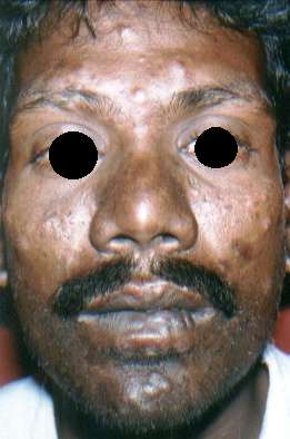
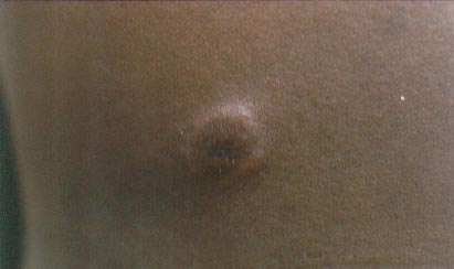
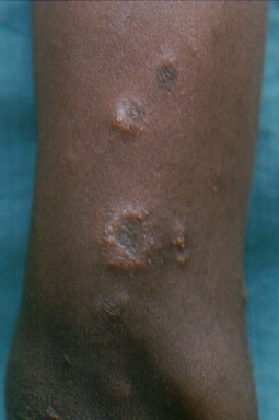
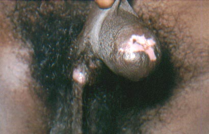
His general physical examination was essentially normal with the exception of multiple, discrete, rubbery, shotty, lymph nodes involving cervical, inguinal and epitrochlear sites. Cutaneous examination revealed discrete, erythematous papules, nodules and annular plaques with mild scaling, over the face(Figure 1), back(Figure 2), and limbs(Figure 3), bilaterally but asymmetrically. Lesions over the eyebrow had resulted in patchy alopecia, though scalp hair was normal. Palms and soles were normal. Split papules were present over the angles of the mouth and papular and nodular lesions were seen over the lips(Figure 1). Examination of the external genitalia showed depigmentation over the prepuce, at the site of the previous ulcer, and a moist, flattened condyloma lata lesion at the peno-scrotal junction (Figure 4).
 |  |
| Figure 1 | Figure 2 |
|---|---|
| Fig.1 Face showing papular and nodular syphilid along with split papules at the angle of mouth | |
| Fig. 2 Nodulo-plaque lesion of secondary syphilis over the back | |
 |  |
| Figure 3 | Figure 4 |
|---|---|
| Fig. 3 Annular secondary syphilis lesions | |
| Fig. 4 Glans penis showing atrophic scar with condylomata lata at penoscrotal junction | |
Laboratory investigations including CBC, blood sugar, renal, and liver function tests were within normal limits. Microscopic examination of the urine and stool were normal. An initial VDRL test for syphilis was negative due to the prozone phenomenon. A repeat test with dilutions was reactive in the dilution of 1:64. His serology for TPHA was reactive. Serological testing for HIV was negative. A skin biopsy from a nodular lesion on the arm showed a patchy infiltrate of lymphomononuclear cells around blood vessels and sweat glands in the dermis. Besides this, epithelioid cell granulomas with plenty of lymphomononuclear cells were also seen in the dermis (Figures 5,6). On the basis of the history, examination findings and serological results, a diagnosis of nodular secondary syphilis was made. The patient received 2.4 million units of benzathine penicillin by the intramuscular route after sensitivity testing and more than 90% of his lesions subsided within ten days. The patient was discharged and asked to come for follow up serological tests after 3 months.
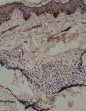 | 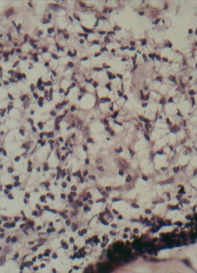 |
| Figure 5 | Figure 6 |
|---|---|
| Fig. 5 Photomicrograph showing patchy infiltrate around capillaries in the dermis (Haematoxylin & Eosin x 100) | |
| Fig. 6 Photomicrograph showing epithelioid cell collection surrounded by lymphomononuclear cells in the dermis (Haematoxylin & Eosin x 400) | |
Discussion
Secondary syphilis usually manifests 3-12 weeks after the appearance of a chancre, and recedes in 4-12 weeks.[1] Skin eruptions develop in 80 to 95 percent of cases. Over 95 percent of the eruptions are macular, maculopapular, or papular lesions that may evolve to become annular, papulosquamous, lenticular, corymbose, or a mixture of these.[1] Nodular and pustular eruptions occur infrequently.[1,2] Mucous membrane lesions are extremely infectious and include condyloma lata, mucous patches and pharyngitis. Our patient presented with papular and nodular lesions, some showing an annular configuration. Though nodular lesions are usually a manifestation of tertiary syphilis, they have also been described in secondary syphilis.[2, 3, 4, 5, 6] In the course of the late secondary stage, nodular and ulcerated lesions may arise as precursors of the tertiary stage.[7] The nodular eruption of secondary syphilis may be localized, but there is a predilection for the face, mucous membranes and palms and soles. Scaling may be present. Though usually no specific pattern is assumed, an annular configuration may be noted.[8]
The differential diagnosis of nodular syphilis includes deep mycoses, leprosy, tuberculosis, sarcoidosis, and lymphoma.[3] In our patient, the results of serological tests and the rapid response to penicillin indicated a correct diagnosis. Histopathological examination in our case showed the presence of an epithelioid granuloma with lymphocytes. The histologic appearance of secondary syphilis is quite variable. Ackerman,[9] in his review on the histologic patterns of secondary syphilis described the following histological patterns - 1) Superficial perivascular dermatitis with epidermal hyperplasia, 2) Superficial and deep perivascular dermatitis (with or without epidermal hyperplasia), and 3) Dense diffuse dermatitis with plasma cell dominant or granulomatous infiltrate (latter divided into those with epithelioid tubercles or with associated fibrosis). The blood vessels typically show dilatation, thickening and hyperplasia of endothelial cells. Thus, granulomas, as seen in our case are an uncommon finding in secondary syphilis and are more a feature of tertiary syphilis. This, together with the fact that our patient had nodular skin lesions highlights the fact that that syphilis may have protean manifestations and the clinical and histologic features of different stages may overlap.[2] The VDRL test usually shows positive results in patients with secondary syphilis and may show negative or low titer positivity in tertiary syphilis.[2]
Nodular syphilis is an uncommon manifestation of secondary syphilis and may go unrecognized. This becomes especially important in the light of the fact that nodular secondary syphilis could be a precursor of tertiary syphilis, a potentially morbid condition. A high degree of clinical suspicion and a careful sexual history, followed by serological testing permits accurate diagnosis, thus facilitating prompt treatment of these cases.
References
1. Sanchez MR. Syphilis, in Dermatology in General Medicine, ed. Fitzpatrick T B, Eisen A Z, Wolff K, et al, 5th edn. McGraw-Hill, New York 1999; 2551-2581.2. Adriaans B. An erythematous nodular eruption. Secondary syphilis. Arch Dermatol 1992;128(7):978-981.
3. Graham WR Jr, Duvic M. Nodular secondary syphilis. Arch Dermatol 1982;118:205-206.
4. Sapra S, Weatherhead L. Extensive nodular secondary syphilis. Arch Dermatol 1989;125:1666-1669.
5. Pavithran K. Nodular secondary syphilis. Int J Dermatol 1991;30:799-800.
6. Papini M, Bettacchi A, Guiducci A. Nodular secondary syphilis. Br J Dermatol 1998;138(4):704-705.
7. Boneschi V, Brambilla L, Bruognolo L. Late secondary syphilis. G Ital Dermatol Venereol 1989;124(5):211-214.
8. Vibhagool C, Raimer SS, Sanchez RL. A nodule on the lip. Nodular secondary syphilis. Arch Dermatol 1996;132(7):822-826.
9. Jeerapet P, Ackerman AB. Histologic patterns of secondary syphilis. Arch Dermatol 1973;107:373-377.
© 2003 Dermatology Online Journal