Navel melanoma: Not always easy to detect, not always difficult to remove
Published Web Location
https://doi.org/10.5070/D32nm0d006Main Content
Navel melanoma: Not always easy to detect, not always difficult to remove
Cristina Mangas, Jorge Romaní, Carlos Muñoz, Jesús Luelmo
Dermatology Online Journal 14 (11): 20
Dermatology Department, Hospital de Sabadell, Parc Taulí, Sabadell, Spain. cmangas@tauli.catAbstract
Melanoma diagnosis can be delayed when the tumor is present in areas that are poorly visualized. We present a peri-umbilical melanoma and discuss the use of Y-plasty in this area.
Introduction
It is well known that melanomas arising in hidden areas such as the umbilicus often suffer from a delayed diagnosis and treatment [1]. Moreover, the umbilicus is a special area that is not always accurately explored but it must be reconstructed, because of its aesthetic relevance for the appearance of the abdomen [2, 3]. Here we report a case of melanoma in the umbilical area in which the tumor was totally resected and the umbilicus was partially reconstructed with a very good aesthetic result.
Case
 |  |
| Figure 1 | Figure 2 |
|---|---|
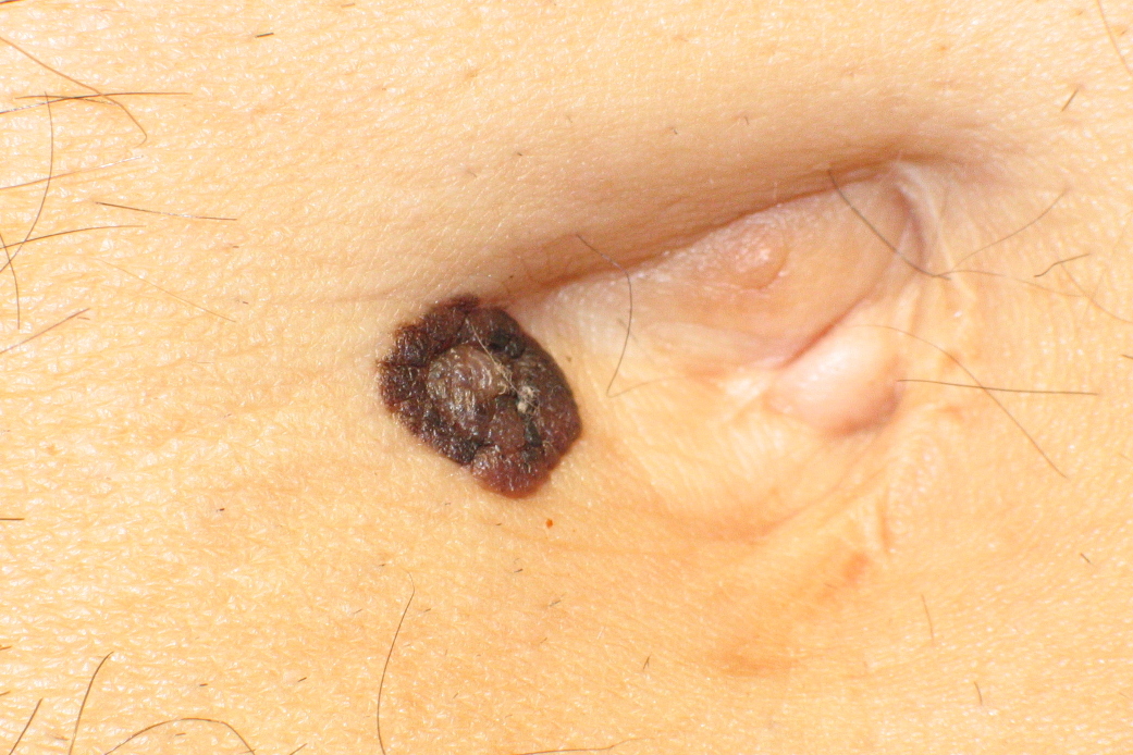
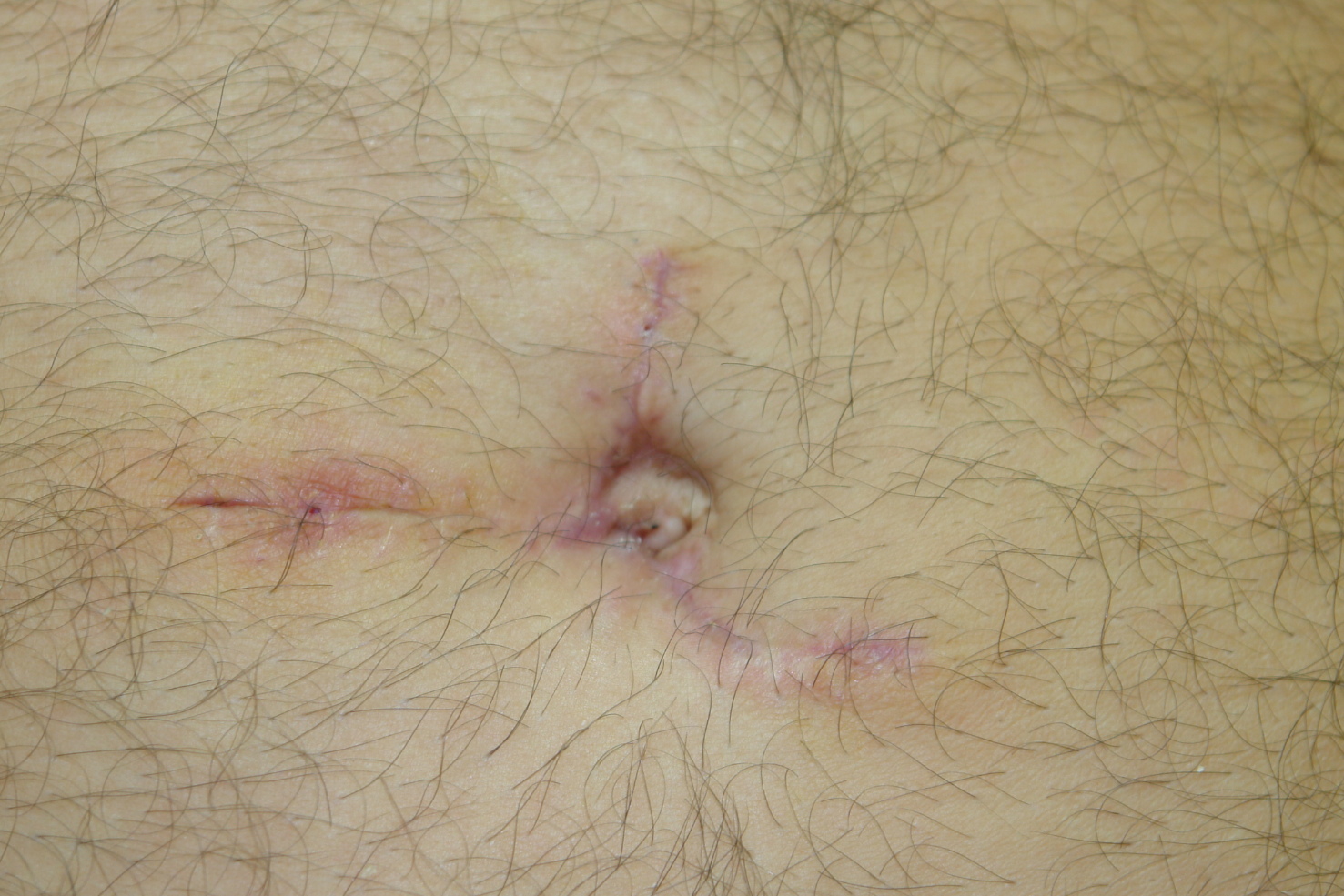
| Figure 1. The patient had this pigmented lesion in the vicinity of the umbilicus Figure 2. Postoperative view of the umbilicus at 1 month | |
A 63-year-old man was referred to our hospital from Primary Care for evaluation of a pigmented lesion in the umbilical area of unknown time of evolution (Fig. 1). He had not noticed the lesion due to the fact that he could not easily see his navel, because of his prominent belly. The patient underwent an inguinal herniorrhaphy 45 years ago with a reconstruction of umbilical area in the same intervention. With the clinical suspicion of melanoma, we excised the lesion with 2 mm margin. Since the tumor was located just beside the umbilicus, we could perform a simple excision and direct closure. Pathological examination showed a superficial spreading malignant melanoma associated to a congenital nevus, Clark's level III, Breslow 0.8 mm, without ulceration or regression. We performed an ultrasonography of the axillar and groin areas due to the difficulty to explore them after the previous surgery and fat deposits. No pathological lymph nodes were observed. In a second procedure, wide local excision of the primary lesion with 1 cm margin was performed. We designed a Y-shaped plasty following the previous lineal scar and 1 cm laterally; we excised deeply around it. Due to the large size of the patient's umbilicus a part of it could be kept attached in place. Both Y branches were reinserted deeply to the remaining umbilicus. We also removed a triangular patch of skin from both sides of the Y branches as a discharge. The lower branch of the Y was partially hidden in the lateral wall of the umbilicus, resembling a natural-appearing scar. After a 6-month follow-up, no local or regional recurrence was observed and a functional and aesthetic scar was achieved (Fig. 2).
Discussion
Melanoma located in the vicinity of the umbilicus does not appear to be very frequent, although its real incidence is difficult to determine due to the lack of statistics about it on the literature [1, 4]. Nevertheless, this particular location for a tumor is of special interest, given the difficulty to explore this area for both the patient and the doctor [2, 3, 5]. The present case illustrates how difficult it can be to detect a tumor in the navel, especially for overweight patients. Moreover, the natural-appearing scar of the navel may make difficult the correct diagnosis of melanocytic lesions in this area. On the other hand, removal of the tumors located in this area often involves a reconstruction of the umbilicus. Several different approaches for reconstruction have been reported to date, based on the use of skin flaps fixed to the rectus aponeurosis in order to obtain a natural-appearing deep cavity at an adequate position [2, 3, 5, 6]. In the case herein reported, it was not necessary to perform a complete reconstruction of the umbilicus, because the patient had a previous large navel that allowed a tumor removal with adequate margins, preserving a part of the original umbilicus. This fact made the reconstruction of the navel easier because we were able to suture and fix the plasty to this remaining umbilicus in the adequate depth and position.
References
1. Thompson JF, Scoyler RA, Kefford RF. Cutaneous melanoma. Lancet. 2005 Feb 19-25;365(9460):687-701. PubMed2. Breuninger H, Zimmermann C. Umbilical reconstruction after excision of melanomas in the area of the umbilicus.Hautarzt. 1996 Apr;47(4):273-5. PubMed
3. Kakudo N, Kusumoto K, Fujimori S, Shimotsuma A, Ogawa Y. Reconstruction of a natural-appearing umbilicus using an island flap: case report. J Plast Reconstr Aesthet Surg 2006;59(9):999-1002. PubMed
4. Rex J, Paradelo C, Mangas C, et al. Single-Institution experience in the management of patients with clinical stage I and II cutaneous melanoma: Results of sentinel lymph node biopsy in 240 cases. Dermatol Surg. 2005 Nov;31(11 Pt 1):1385-93. PubMed
5. Castillo PF, Sepúlveda CA, Prado AC, Troncoso AL, Villamán JJ. Umbilical reinsertion in abdominoplasty: technique using deepithelialized skin flaps. Aesth Plast Surg 2007;31(5):519-20. PubMed
6. Onishi K, Yang YL, Maruyama Y. A new lunch box-type method in umbilical reconstruction. Ann Plast Surg 1995;35(6):654-6. PubMed
© 2008 Dermatology Online Journal