Bullous lichen planus of the glans penis
Published Web Location
https://doi.org/10.5070/D327m350xnMain Content
Bullous lichen planus of the glans penis
Kaliaperumal Karthikeyan, B Jeevankumar, and Devinder M Thappa
Dermatology Online Journal 9 (5): 31
Department of Dermatology and STD, JIPMER, Pondicherry, India. dmthappa@jipmer.edu
Lichen planus is a papulosquamous disorder generally limited to flexures, lower extremities, genitalia, and oral mucosa [1]. It is a distinct clinical entity with characteristic color and morphology that manifests characteristic patterns of evolution. Microscopic features, similar to the gross morphology, are diagnostic. Although 25 percent of lichen-planus patients have involvement of the genitalia, isolated involvement of the male external genitalia is rare[2] . We herewith report a case of bullous lichen planus limited to male external genitalia.
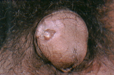
A 45-year-old laborer presented with a penile blister of 3 days' duration. The blister developed within an asymptomatic purple lesion on the glans penis of 5 months' duration. There was no preceding history of trauma. He had not taken any medications prior to the onset of the lesion. The patient denied a history of extramarital sexual contact. Physical examination revealed a solitary 1-cm by 0.75-cm tense bulla overlying an annular violaceous plaque on the dorsal surface of the glans penis (Fig.1). The bulla was filled with serosanguinous fluid. Regional inguinal lymph nodes were not enlarged. The skin and oral mucosa were otherwise normal.
|
| Figure 1 |
|---|
| A tense bulla overlying an annular violaceous plaque on the dorsal surface of the glans penis |
The patient's routine hemogram and biochemistry were within normal limits. His VDRL and HIV serology were nonreactive. Biopsy from the edge of the lesion showed hypergranulosis, hydropic degeneration of the basal cell layer and a bandlike infiltrate composed of mainly lymphocytes in the papillary dermis, together with melanin incontinence. Subepidermal clefts were seen at a few sites in the sections (Figs. 2, 3). Civatte bodies were seen in the lower epidermis and papillary dermis. The patient was diagnosed to have bullous lichen planus of glans penis and was put on betamethasone valerate (0.1 %) cream. The patient responded well to topical steroid therapy, with disappearance of the bulla in a week.

|

|
| Figure 2 | Figure 3 |
|---|---|
| Photomicrograph showing hypergranulosis, hydropic degeneration of the basal cell layer, and a bandlike infiltrate in the papillary dermis, hugging the epidermis (Fig. 2)(Hematoxylin & Eosin x 40) | |
| Higher magnification photomicrograph showing a sub-epidermal cleft with mixed inflammatory infiltrate, with melanin incontinence, in the dermis (Fig. 3)(Hematoxylin & Eosin x 200) | |
Lichen planus of the male external genitalia commonly involves the glans penis, the lesions being typically annular.[2] Violaceous flat-topped papules similar to lesions elsewhere in the skin can also occur on the glans and shaft of the penis. The lesions may develop as arcuate groupings of individual papules that evolve into rings of clustered papules with central clearing. Fine white streaks are usually visible on the surface of the lesions (Wickham striae). Rarely, an erosive lesion can occur on glans penis. Solitary lichen planus of the glans penis alone is very rare. [3] Altman and Perry [4] reviewed the records of 307 patients with lichen planus seen at the Mayo clinic from 1950 to 1954 and reported only one case with involvement of the genitalia alone.
Our patient developed the bullous lesion over preexisting lichen planus on the penis, a feature not reported in the literature. Bullous lichen planus is rare and is the result of extreme liquefaction and vacuolation of the basal cell layer [5]. Occurrence of bullous lichen planus over the glans penis should be differentiated from other conditions, such as fixed-drug eruption. Fixed-drug eruption is characteristically preceded by the intake of the responsible drug, and there is a history of recurrence of similar lesions at the same site. It was intriguing to note the isolated occurrence of the rare bullous lichen planus of the glans penis in our case.
References
1. Black MM. Lichen planus and lichenoid disorders. In: Champion RH, Burton JL, Burns DA, Breathnach SM, eds. Textbook of Dermatology, Vol. 3, 6th ed. Oxford: Blackwell Science 1998, p 1899-1926.2. Daoud MS, Pittelkow MR. Lichen planus, In: Freedberg IM, Eisen AZ, Wolff K, Austen KF, Goldsmith LA, Katz SI, Fitzpatrick TB, eds. Dermatology in General Medicine, Vol.1, 5th ed. New York: Mc Graw-Hill 1999 p 561-576.
3. Alinovi A, Barella PA, Benoldi D. Erosive lichen planus involving the glans penis alone. Int J Dermatol 1983; 22: 37-38.
4. Altman J, Perry HO. The variations and course of lichen Planus. Arch Dermatol 1961; 84: 179-191.
5. Gawkrodger DJ, Stavropoulous PG, Mclaren KM, Buxton PK. Bullous lichen planus and lichen planus pemphigoides: Clinico- pathological comparisons. Clin Exp Dermatol 1989; 14: 150-153.
© 2003 Dermatology Online Journal