Bowen disease over photoprotected site in an Indian male
Published Web Location
https://doi.org/10.5070/D323f586bkMain Content
Bowen disease over photoprotected site in an Indian male
Suruchi Gupta MD, Nutan MD, Sunil Dogra MD DNB, Amrinder J Kanwar MD
Dermatology Online Journal 15 (10): 16
Post Graduate Institute Of Medical Education and Research, Chandigarh, India. docnutan66@gmail.comAbstract
Bowen Disease is squamous cell carcinoma in situ in which the basement membrane is intact on histopathology. Lesions are usually solitary but may be multiple in 10-20 percent of cases. About three-quarters of these lesions are situated on the lower limb. It typically presents as an erythematous enlarging plaque having irregular borders with scaling and crusting. Our patient presented with a lesion on the chest that was not sun exposed thus leading to a diagnostic dilemma.
Case synopsis
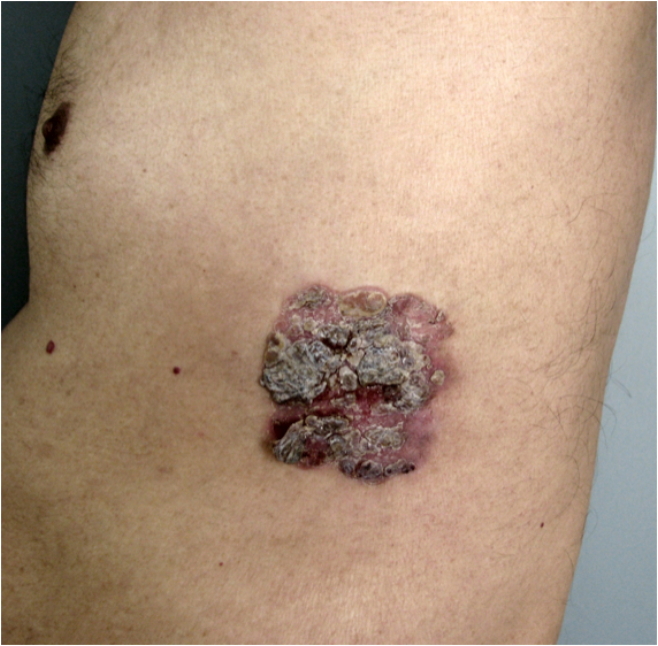 |  |
| Figure 1 | Figure 2 |
|---|---|
| Figure 1. A well-defined erythematous plaque on the lateral thoracic wall with scaling and crusting Figure 2. Windswept appearance with keratinocyte dysplasia and intact basement membrane (H&E, x100) | |
A 58-year-old North Indian male presented to dermatology clinic with the complaint of a gradually enlarging erythematous, infiltrated, crusted plaque over the left side of his abdomen for the past eight years (Fig. 1). It started as a small papule and gradually increased in size to become a persistent large plaque. The overlying crust could be removed by scraping, leaving behind a raw surface. The plaque was occasionally itchy and there was a history of pain on direct pressure. The patient had been given treatment in the form of topical corticosteroids, tar preparations, and salicylic acid combinations without relief. The patient was a teacher and there is no history of sun exposure of the chest or back. The patient was otherwise maintaining good health and there was no history suggestive of chronic arsenic ingestion or exposure to fungicides or pesticides. The patient had no history of underlying medical problems or a history of malignancies. In spite of the peculiar location, the clinical diagnosis was Bowen disease. Histopathology was performed which was confirmatory (Fig. 2). The patient was started on topical Imiquimod 5 percent ointment applied on alternate days, which produced complete clearance in two months. However, there was recurrence at three months follow up and he was advised to have excision.
Discussion
Bowen disease is a squamous cell carcinoma in situ with the potential for significant lateral spread [1]. Earlier it was thought to be a paraneoplastic condition, but in large population based cohorts the associations with underlying malignancies was ruled out [2, 3]. It is most commonly seen in elderly light skinned people without any sex predilection. The incidence is around 142 in 100,000 population in a study from Hawaii in 1994 [4]. The causative factors implicated are excessive sun exposure, chronic arsenic poisoning, genetic factors, trauma, other carcinogenic chemicals and X ray exposure. Some studies have documented the association of human papilloma virus type 16 and type 2 with Bowen disease.
The condition usually presents as a single plaque in mucocutaneous sites in the head and neck area. The appearance is of an erythematous, infiltrated, scaly, crusted, fissured plaque that can be commonly confused with psoriasis, nummular eczema, and actinic keratosis. Sometimes the lesion may be pigmented and can be confused with melanoma. Other entities in the differential diagnosis to be considered are basal cell carcinoma, extra mammary Paget disease, plaque-type psoriasis and tinea corporis.
Histopathology is characterized by a full-thickness atypia of the epidermis, with loss of the normal maturation of its components. Keratinocytes are atypical and disorderly, often described as having a windblown appearance. Although the basal cell layer is intact, extension of keratinocyte atypia down the follicular epithelium is seen. The pagetoid cells are large pale keratinocytes with abundant ground glass cytoplasm, which are distributed haphazardly throughout the epidermis.
Various medical modalities have been tried in Bowen disease including 5 percent flourouracil, 5 percent imiquimod, grenz ray radiation and photodynamic therapy [5]. Amongst surgical choices simple excision with wide margins, Mohs micrographic surgery, carbon dioxide laser, cryotherapy and curettage with electrodessication have been used with good success.
References
1. http://emedicine.medscape.com/article/1100113-overview2. Arbesman H, Ransohoff DF. Is Bowen's disease a predictor for the development of internal malignancy?A methodological critique of the literature. JAMA. 1987 Jan 23-30; 257(4): 516-518. [PubMed]
3. Jaeger AB, Gramkow A, Hjalgrim H, Melbye M, Frisch M. Bowen disease and risk of subsequent malignant neoplasms: a population-based cohort study of 1147 patients. Arch Dermatol. 1999 Jul; 135(7): 790-3. [PubMed]
4. Reizner GT, Chuang TY, Elpern DJ, Stone JL, Farmer ER. Bowen's disease (squamous cell carcinoma in situ) in Kauai, Hawaii. A population-based incidence report. J Am Acad Dermatol. 1994 Oct; 31(4): 596-600. [PubMed]
5. Cox NH, Eedy DJ, Morton CA. Guidelines for management of Bowen's disease: 2006 update. Br J Dermatol. 2007 Jan; 156(1): 11-21. [PubMed]
© 2009 Dermatology Online Journal