Lichen planus pemphigoides: a case report and review of the literature
Published Web Location
https://doi.org/10.5070/D31xs8j0mrMain Content
Lichen planus pemphigoides: a case report and review of the literature
Mandy S Harting MD, Sylvia Hsu MD
Dermatology Online Journal 12 (4): 10
Department of Dermatology, Baylor College of Medicine, Houston, Texas. shsu@bcm.edu Lichen planus pemphigoides (LPP) is thought to be a rare variant of bullous pemphigoid (BP). It is characterized by bullous lesions arising on lichen planus papules and on clinically uninvolved skin. It is reported that lichen planus pemphigoides is induced by numerous medications including the following: cinnarizine, captopril, ramipril, simvastatin, PUVA, and antituberculous medications [1, 2, 3, 4]. Some cases of LPP demonstrate overlapping characteristics with paraneoplastic pemphigus (PNP) and have been associated with internal malignancy. One report by Hamada et al. discusses a case of LPP associated with colon adenocarcinoma and numerous keratoacanthomas [6].
Clinical synopsis
A 44-year-old woman with obesity, diabetes mellitus, and hypertension presented with a 4-month history of multiple pruritic blisters on her lower extremities. The blisters mainly occurred on her right lower extremity, and there was no truncal involvement. She also complained of several pruritic plaques without blisters on her bilateral lower extremities that had been present for approximately 1 year. She had not started any new medications for many years.
On examination, she had numerous lichenified plaques, papules, and excoriations on both lower extremities. Within these plaques and also surrounding these plaques, she had numerous tense bullae. The skin lesions were completely restricted to her lower extremities. Besides a moderately uncontrolled glucose, all of her remaining laboratory values, including tests for hepatitis B and C, were negative or within normal limits.
Two skin specimens were examined. The first biopsy was taken from one of the plaques and showed lichenoid dermatitis consistent with lichen planus. The second skin biopsy was performed on the edge one of one of the bullae. The specimen showed a subepidermal split with eosinophils. The direct immunofluorescence demonstrated C3 and IgG in a linear pattern at the basement membrane zone. These features were consistent with bullous pemphigoid.
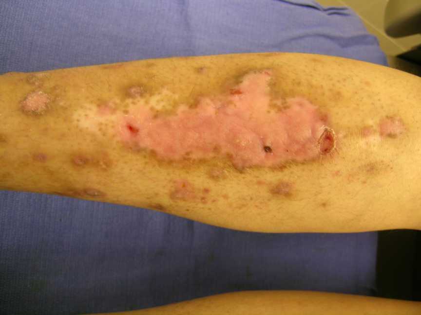 |
| Figure 1 |
|---|
| Lichenified plaques on the shins after 2-week treatment with clobetasol ointment BID |
Based on our clinical findings in conjunction with the pathologic specimens, a diagnosis of lichen planus pemphioides was made. We did not believe that our patient was a candidate for systemic steroids in part because of her poorly controlled diabetes mellitus along with her relatively mild disease distribution. Therefore, we elected to treat her with topical clobetasol ointment. Our patient responded well to potent topical steroids, and she has not had new bullae after using the topical steroid (Fig. 1).
Comment
Compared to bullous pemphigoid, lichen planus pemphigoides is thought to affect a younger age group and have a less severe clinical course. Two recent reviews revealed the mean age of onset of LPP to be 35 and 44 years; bullous pemphigoid usually presents at an older age. When comparing the location of bullae in LPP versus BP, the lesions tend to occur on the limbs and trunk, respectively [7]. Lichen planus pemphigoides responds well to standard therapies, with systemic steroids being the most effective treatment for extensive disease [1, 5]. Many other treatment choices have been used besides topical and systemic steroids. Other treatments include tetracycline and nicotinamide, isotretinoin, dapsone, and immunosuppressive drugs [1].
Many cases of LPP in the literature have demonstrated IgG autoantibodies to either one or both the 180 kd pemphigoid antigen (BPAg2, type-XVII collagen) and the 230 kd pemphigoid antigen (BPAg1) [1, 5, 6, 8]. Some case reports even describe a pattern more consistent with paraneoplastic pemphigus [6]. It has been proposed that damage to the basal cells in LP or damage due to other regimens such as PUVA unmask hidden antigenic determinants, or create new antigens, leading to antibody formation and induction of BP [1, 4].
Lichen planus pemphigoides is a rare clinical variant of bullous pemphigoid that is important to recognize because its course tends to be much more indolent, and it responds well to treatment. As clinicians, we must be aware of drug-induced etiologies and be mindful that some cases have been associated with internal malignancies. Therefore, in patients with LPP, it is extremely important to elicit a careful history, perform a thorough examination, and advise our patients to have all standard cancer screenings recommended.
References
1. Demircay Z, Baykal C, Demirkesen C. Lichen planus pemphigoides: report of two cases. Int J Dermatol 2001;40(12):757-760.2. Ogg GS, Bhogal BS, Hashimoto T, Coleman R, Barker JN. Ramipril-associated lichen planus pemphigoides. Br J Dermatol 1997; 136(3):412-414.
3. Stoebner PE, Michot C, Ligeron C, Durand L, Meynadier J, Meunier L. Simvastatin-induced lichen planus pemphigoides. Ann Dermatol Venereol 2003;130(2 Pt 1):187-190.
4. Kuramoto N, Kishimoto S, Shibagaki R, Yasuno H. PUVA-induced lichen planus pemphigoides. Br J Dermatol 2000;142(3):509-512.
5. Sakuma-Oyama Y, Powell AM, Albert S, Oyama N, Bhogal BS, Black NM. Lichen planus pemphigoides evolving into pemphigoid nodularis. Clin Exper Dermatol 2004 ;28(6):613-616.
6. Hamada T, Fujimoto W, Okazaki F, Asagoe K, Arata J, Iwatsuki K. Lichen planus pemphigoides and multiple keratoacanthomas associated with colon adenocarcinoma. Br J Dermatol 2004;151(1):252-254.
7. Swale, Black, Bhogal. Lichen planus pemphigoides: two case reports. Clin Exper Dermatol 1998;23(3):132-135.
8. Hsu S, Ghohestani RF, Uitto J. Lichen planus pemphigoides with IgG autoantibodies to the 180kd bullous pemphigoid antigen (type XVII collagen). J Am Acad Dermatol 2000;42(1 Pt 1):136-141.
© 2006 Dermatology Online Journal