Pyogenic granuloma on the hand subsequent to friction blister in a hand surgeon
Published Web Location
https://doi.org/10.5070/D30zv279dwMain Content
Pyogenic granuloma on the hand subsequent to friction blister in a hand surgeon
Sezai Sasmaz MD1, Ahmet Karaoguz MD2, Murat Uzel MD2, Y Kenan Coban, MD3
Dermatology Online Journal 12 (3): 22
1. KSU Faculty of Medicine, Department of Dermatology, Kahramanmaras-Turkey. drsasmaz@gmail.com2. KSU Faculty of Medicine, Department of Orthopedic Surgery, Kahramanmaras-Turkey
3. KSU Faculty of Medicine, Department of Plastic and Reconstructive Surgery, Kahramanmaras-Turkey
Abstract
A case of pyogenic granuloma in a 62-year-old hand surgeon secondary to friction blister is reported. He developed a fast growing, hypertrophic inflammatory papule consisting of exceptionally richly vascularized granular tissue. This dusky red lesion was located on the thenar part of the hand. We find no prior report of PG attributed to friction blister.
Friction blisters (FBs) occur frequently, especially in vigorously active populations. A FB is a soft pocket of raised skin filled with clear fluid. It tends to occur in areas of thick adherent stratum corneum such as palms and soles. Studies using rubbing techniques show that FBs result from frictional forces that mechanically separate epidermal cells at level of the stratum spinosum. The magnitude of frictional forces and the number of times that an object cycles across the skin determine the probability of blister development; the higher the frictional forces, the fewer the cycles necessary to produce a blister [1, 2].
Most FBs heal on their own in a few days. A new layer of skin forms beneath the blister, and eventually the blistered skin peels away. If pressure or friction continues in the same area, the blister may last two weeks or longer. Continued friction may rub away the delicate top skin layer, and the blister may break open, ooze fluid and run the risk of becoming infected. Impetigo may become a serious complication of FB with resulting cellulitis and sepsis [1, 2]. We present a unique case of pyogenic granuloma (PG) as the other complication of FB.
Clinical synopsis
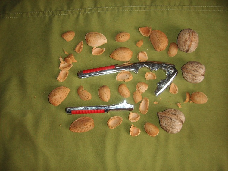
A 62-year-old hand surgeon was seen for a blister on the thenar area of the right palm attributed to the use of a nut cracker (Fig. 1). The patient presented with a tender, small pocket of puffy, raised skin containing clear fluid. The clinical diagnosis was FB, and it was drained to releive the discomfort attributed to the tension. The roof of the blister was left intact to serve as a wound dressing. The patient was instructed to apply an antimicrobial ointment daily to the treated area for a few days.
 |  |
| Figure 1 | Figure 2 |
|---|---|
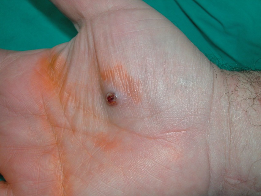
| Figure 1. The nut cracker Figure 2. Dusky red, vascular lesion on the thenar area of the right palm | |
On his return visit after 1 month, we observed an indolent, dusky red, flattened, flesh-like oval mass of 8 mm in diameter at the previous site of the FB (Fig. 2). The lesion appeared to be erupting through a gap in the skin. The patient stated that the lesion had developed approximately 15 days after the development of FB, had grown rapidly and bled easily when traumatized. There was no history of additional injury or infection.
The clinical diagnosis was pyogenic granuloma (PG). The lesion was destroyed by electrosurgery and curettage leaving no specimen for histopathologic confirmation. The treated site healed with a small scar in 10 days. At 20 months followup, there is no evidence of recurrence.
Discussion
PG is a rapidly developing vascular lesion that often arises at sites of minor trauma, such as on the face, fingers, toes, and trunk; it has a clear preponderance for developing in highly vascular tissues [3]. The majority of PGs appear on the head and neck but a substantial minority (12-37 %) involves the hands and forearms [4]. It varies in size from a few millimeters to 2 cm and it commonly occurs in childhood or in early adult life [3, 4, 5, 6]. The term PG is a misnomer. Originally, these lesions were thought to be caused by bacterial infection; however, the etiology has not been determined. The histopathologic appearance is fairly characteristic; the lesion is, in fact, a lobular capillary hemangioma [5]. Most authors believe that this vascular proliferation is a reactive and hyperplastic condition, rather than a true neoplasm [6].
The clinical diagnosis of PG is usually easy to make. Sudden onset and rapid enlargement are common. Bleeding is typical.
A nitric oxide synthase-dependent mechanism is thought to contribute to angiogenesis and the rapid growth of PGs. The patients or their parents are frequently concerned that the rapid growth and bleeding may indicate a malignancy. Recognition of PG as a clinically polypoid or exophytic circumscribed lesion is of importance to the clinician because this feature distinguishes PG from most malignant vascular tumors [5]. Vascular cancers such as Kaposi sarcoma occasionally occur as solitary friable nodules. These cancers typically grow slowly, however, and cause purple discoloration around the lesion [7]. The other skin conditions to consider in the differential diagnosis are squamous cell carcinoma and amelanotic melanoma [5, 7]. Squamous cell carcinoma can develop as a rapidly growing reddish nodule. It tends to have scale or crust on the surface, unlike the lesion seen in our case. As for amelanotic melanoma, it can grow fairly quickly, may bleed easily, and can lack brown pigmentation [7]. Nevertheless, it would be very unusual for a melanoma to attain the size of 8 mm in diameter after only 2 weeks.
In the case presented here, a PG occurred subsequent and specifically related to an FB. The lesion was a single, painless, reddish mass, and located on thenar part of the right palm. These characteristics were in accordance with the general pattern of PG. However, the patient was a hand surgeon and older than the more commonly reported second decade of life. Instead of erythematous papule, a flattened, flesh-like mass was seen. The clinical development was not slow but rapid; over a 2-week period the lesion increased in size until it reached 8 mm diameter.
Treatment of PGs includes surgical excision, curettage, electrical cauterization, shaving with cauterization, flashlamp-pumped pulsed dye laser ablation, low-dose external beam radiation, and silver nitrate cauterization [3, 4]. Electrosurgical treatment is the treatment of choice for solitary lesions. To provide an adequate cure rate, all vascular granulation tissue must be removed or cauterized [5, 8]. Recurrence is more likely when they are incompletely removed, but also possible after apparently complete removal [5].
PGs can appear within an existing capillary malformation (port-wine stain), but most commonly there is no history of a preexisting dermatologic condition [3]. Development of PG has not been reported previously as a complication of FB.
References
1. Knapik JJ, Reynolds KL, Duplantis KL, Jones BH. Friction blisters. Pathophysiology, prevention and treatment. Sports Med. 1995 Sep;20(3):136-147. PubMed2. Kuljit C, Lambert WC, Schwartz RA. Friction blisters. Available from URL: http://www.emedicine.com/derm/topic.161.htm (Accessed 2005 June 14).
3. Grevelink SV, Mulliken JB. Vascular anomalies and tumors of skin and subcutaneous tissues. In: Freedberg IM, Eisen AZ, Wolff K, et al., eds. Fitzpatrick's dermatology in general medicine, 6th ed. New York: McGraw-Hill, 2003:1002-19.
4. Quitkin HM, Rosenwasser MP, Strauch RJ. The efficacy of silver nitrate cauterization for pyogenic granuloma of the hand. J Hand Surg. 2003 May;28(3):435-8. PubMed
5. Crowe MA, Steinberg B. Pyogenic granuloma. Available from URL: http://www.emedicine.com/ped/topic1244.htm (Accessed 2005 June 14).
6. Requena L, Sangueza OP. Cutaneous vascular proliferations. Part II. Hyperplasias and benign neoplasms. J Am Acad Dermatol. 1997 Dec;37(6):887-919. PubMed
7. Levine N. Red lesion on the hand. Irregular nodule grows quickly and bleeds easily when exposed to minor trauma. Geriatrics. 2001 Aug;56(8):23. PubMed
8. Hainer BL. Electrosurgery for the skin. Am Fam Physician 2002 Oct 1;66(7):1259-66. PubMed
© 2006 Dermatology Online Journal