Unilateral non-pigmenting fixed drug eruption associated with cotrimoxazole
Published Web Location
https://doi.org/10.5070/D30qk2338pMain Content
Unilateral non-pigmenting fixed drug eruption associated with cotrimoxazole
Abbas Rasi MD1, Alireza Khatami MD2
Dermatology Online Journal 12 (6): 12
1. Department of Dermatology, Hazrat-e-Rasoul Hospital, Iran University of Medical Sciences, Tehran, Iran. abbasrasi@yahoo.com2. Center for Research and Training in Skin Diseases and Leprosy, Tehran
Abstract
The pathogenesis of fixed drug eruption (FDE) is still unknown. One of the most common associations of FDE is the use of sulfonamides. Cotrimoxazole (trimethoprim with sulfamethoxazole) is one of the most commonly prescribed sulfonamide drugs. Non-pigmenting FDE (NPFDE) is a a relatively rare condition and only a few cases have been reported. We describe a case of unilateral NPFDE in a 45-year-old man whose lesions were on his right leg and foot as well as his ipsilateral penile skin. Cotrimoxazole was suspected as the offending drug and its role was confirmed by an oral challenge test.
Fixed drug eruption (FDE) is a cutaneous drug reaction characterized by the recurrent appearance of skin lesions at the same site or sites each time the responsible drug is administered. The eruption appears usually within 30 minutes to 8 hours after drug exposure. Fixed drug eruption lesions are characteristically sharply marginated, round or oval pruritic plaques of erythema and edema. Fixed drug eruption typically resolves leaving behind discrete, postinflammatory hyperpigmented macules or patches. The diagnostic hallmark is its recurrence at the previously affected sites. Fixed drug eruption can be attributed to a variety of drugs. Sulfonamides are the most commonly implicated. Cotrimoxazole (a combination of trimethoprim and sulfamethoxazole), is widely used for treatment of infectious diseases and is the most commonly implicated drug. Oral provocation is still the gold-standard method for establishing the offending agent, but topical provocation would be a safer first step [1, 2, 3, 4, 5].
Clinical synopsis
A 45-year-old man who had been treated with cotrimoxazole for sinusitis presented with a sudden onset of erythematous plaques and patches. He had a 3-year history of sinusitis for which he had taken cotrimoxazole intermittently. Because of fatigue, malaise, and aggravation of his sinusitis symptoms, he took a single dose of 800/160 mg cotrimoxazole 2 days prior to admission to the dermatology clinic. He mentioned that about 6 hours after ingesting the drug, round, red, and slightly tender skin lesions appeared on his right leg, right foot, and the right side of his penis. The patient reported similar cutaneous eruptions on the same body areas after cotrimoxazole intake about 1 and 3 years before his current presentation. He otherwise had normal health and had no history of allergies. On physical examination there were discrete, round, red-to-violaceous plaques and patches in a linear distribution on patient's right leg, right foot, and right side of the penis.
 |  |
| Figure 1 | Figure 2 |
|---|---|
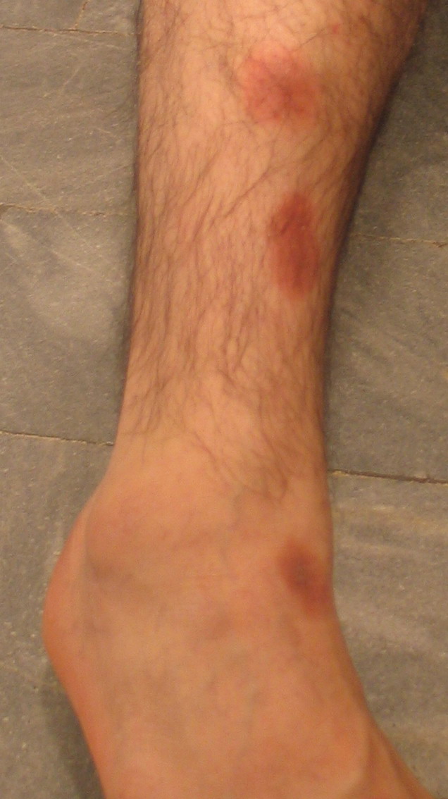
| Figure 1. Recurrent, well-defined, erythematous patches on the skin of a patient for whom the primary diagnosis of unilateral
non-pigmenting FDE was suggested. Lesions on his right lower extremity. Figure 2. Skin lesions on right side of his penis. After oral challenge test the diagnosis of unilateral NPFDE from cotrimoxazole confirmed. | |
The eruption was diagnosed as FDE and cotrimoxazole treatment was discontinued. He was treated with topical 0.05 percent clobetasol propionate ointment. The eruption cleared completely without residual pigmentation in 2 weeks. The patient refused to undergo closed patch testing to determine the causative agent. Oral challenge test was carried out after 3 weeks, using one-half of a single pediatric cotrimoxazole tablet (i.e., 50 mg sulfamethoxazole + 10 mg trimethoprim). After 30 minutes, erythematous patches re-appeared at the previously involved areas, accompanied with itching and burning sensation, confirming that the patient had NPFDE associated with cotrimoxazole. During a 1-year followup period, the patient was not re-exposed to cotrimoxazole and he did not have any skin lesions.
Discussion
FDE was first described by Bourns in 1889, but Brocq then coined the name some years later [1]. Abramovitz and Noun proposed the concept of non-pigmenting FDE (NPFDE). Some 50 years later Shelly and Shelly described a distinctive type of NPFDE that consisted of symmetric, tender, large, erythematous plaques [6]. Since then, there have been reports of NPFDE associated with ephedrine, pseudo-ephedrine, tetrahydrozoline hydrochloride, piroxicam, thiopental, radiopaque contrast media iothamalate, diflunisal, arsphenamine, paracetamol, intra-articular triamcinolone acetonide, and eperisone hydrochloride [3, 6].
The pathogenesis of FDE remains undetermined. However, results of a recent study suggested that T cells residing in the epidermis contribute to tissue injury [7]. Shiohara et al. and Teraki et al. demonstrated that CD8+ T cells are enriched in the lesions of FDE, suggesting that improper T-cell activation is responsible for epidermal tissue injury [8, 9]. Although the pathogenesis of FDE is still unclear, the major classes of drugs associated with this disorder are well recognized [3].
There are no definitive laboratory tests to confirm the diagnosis of FDE. Oral provocation with the suspected agent is the only reliable method in most cases [10]. The clinical pattern of FDE may provide useful information to determine the most likely causative drug, especially when the details of the drugs to which the patient has been exposed are known [4]. Fixed drug eruption is characterized by well-circumscribed erythematous patches and plaques, occasionally associated with bulla formation. Characteristically, FDE eruptions recur at the same sites with repeated exposure to the offending drug. The lips, genitalia, and sacral area are favored involvement sites, however FDE lesions can appear on any part of the body [3].
Cotrimoxazole is mentioned as the most frequently associated drug in several studies. In our case, the onset of the eruption was 6 hours after cotrimoxazole exposure. Following resolution of the lesions, recurrence of the non-pigmented lesions with re-administration of the drug strongly suggests the diagnosis of cotrimoxazole-induced NPFDE. Trimethoprim has been implicated in linear FDE, but unilateral NPFDE from to cotrimoxazole has not been reported [11]. Although the patient refused a patch test with cotrimoxazole, the result of oral challenge with that drug was positive confirming its role.
We believe this is the first reported case of unilateral NPFDE associated with cotrimoxazole. Recognition of this rare clinical entity is important because the diagnosis of unusual FDE presentations, such as linear NPFDE following exposure to cotrimoxazole, may be missed by physicians.
References
1. Al-Mutairi N, Al-Fouzan A, Nour-Eldin O. fix drug eruption due to Influenza Vaccine. J Cutan Med Surg 2004 Jan-Feb;8(1):16-8. PubMed2. Heikkila H, Timonen K, Stubb S. Fix drug eruption due to Fluconazole. J Am Acad Dermatol 2000 May;42(5 Pt 2):883-4. PubMed
3. Breathnach SM. Drug reactions. In: Burns T, Breathnach SM, Cox N, Griffits C (editors). Rook's Textbook of Dermatology. 7th edition. Oxford. Blackwell Science: 2004; 73:28.
4. Ozkaya-Bayazit E, Bayazit H, Ozarmagan G. Drug related clinical pattern in fixed drug eruption. Eur J Dermatol 2000 Jun;10(4):288-91. PubMed
5. Mahboob A, Haroon TS. Drugs causing fixed eruptions: a study of 450 cases. Int J Dermatol 1998 Nov;37(11):833-8. PubMed
6. Shelley WB, Shelley ED. Nonpigmenting fix drug eruption as a distinctive reaction pattern: examples caused by sensitivity to pseudoephedrine hydrochloride and tetrahydrozoline. J Am Acad Dermatol 1987 Sep;17(3):403-7. PubMed
7. Shiohara T, Mizukawa Y, Teraki Y. Pathophysiology of fix drug eruption:the role of skin- resident T cells. Curr Opin Allergy Clin Immunol 2002 Aug;2(4):317-23. PubMed
8. Shiohara T, Nicoloff BG, Sagawa Y, Gomi T, Nagashima M. fix drug eruption. Expression of epidermal keratinocyte intercellular adhesion molecule-1 (ICAM-1). Arc Dermatol 1989 Oct;125(10):1371-6. PubMed
9. Teraki Y,Moriya N,Shiohara T. Drug-induced expression of intercellular adhesion molecule-1 on lesional keratinocytes in fix drug reaction. Am J Pathol 1994 Sep;145(3):550-60. PubMed
10. Stubb S, Heikkila H, Kauppinen K. Cutaneous reactions to drugs:a series of inpatients during a five-year study.Acta Derm Venereol 1994 Jul;74(4):289-91;74:289-291. PubMed
11. Ozkaya-Bayazit E, Baykal C. Trimethoprim-induced linear fixed drug eruption. Br J Dermatol 1997 Dec;137(6):1028-9. PubMed
© 2006 Dermatology Online Journal