Alopecia areata of eyelashes: A subset of alopecia areata
Published Web Location
https://doi.org/10.5070/D307t0791xMain Content
Alopecia areata of eyelashes: A subset of alopecia areata
Naga Prasad Grandhe MD, and Amrinder Jit Kanwar MD
Dermatology Online Journal 10 (2): 13
Department of Dermatology, Venereology and Leprology, Postgraduate Institute of Medical Education and Research, Chandigarh,
India. ajkanwar@sify.com
Alopecia areata (AA) is an autoimmune disease characterized by unpredictable, usually patchy, nonscarring hair loss. Although it most commonly affects the scalp, it can involve any hair-bearing area on the body. Loss of eyelashes and eyebrows can occur with severe forms of AA, but presentation of AA with exclusive involvement of eyelashes is extremely rare [1, 2]. We present a child who had bilateral patchy loss of eyelashes resulting from AA.
Clinical synopsis
|
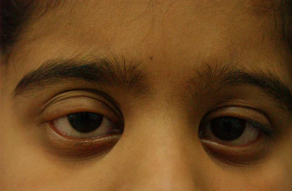
| Figure 1 |
|---|
| Alopecia areata of eyelashes with patchy loss of eyelashes of upper and lower eyelids bilaterally. |
A 9-year-old female child presented with an asymptomatic, gradual loss of eyelashes for 6 months. She had no history of acute illness prior to the onset of the hair loss. She denied symptoms of blepharitis, and no history of any systemic medication was elicited. There was no past or present history of a psychiatric disorder. There were neither signs nor history suggestive of atopy or thyroid disease, and there was no family history of these diseases.
On examination, there was patchy loss of eyelashes involving upper and lower eyelids of both the eyes (Fig. 1). There were no signs of inflammation over lid margins. No other patches of hair loss could be identified over the scalp or rest of the body. Examination of her nails and mucosa did not reveal any abnormality. Secondary lid-margin pathology was ruled out through a slit-lamp examination. Her visual acuity and intraocular pressure were normal. After consideration of her findings, a diagnosis of alopecia areata confined to the eyelids was considered and the child was started on systemic steroids (oral minipulse betamethasone tab 2.5 mg twice a week). After 3 months of this therapy significant eyelash regrowth was observed.
Discussion
The prevalence of AA among the dermatology outpatients is 2 percent [3]. The majority of these patients (60 %) present with the first patch of AA before age 20 [4, 5]. Long-term prognosis of AA is relatively poor in those with an onset at younger age [6]. AA is categorized into three forms according to the extent of involvement: partial alopecia, alopecia totalis, and alopecia universalis. In 10 percent of cases of partial alopecia, the lesions are located at the sites other than the scalp [7]. Few cases of AA confined to the eyelashes are reported in the literature [1, 2]. According to Duke-Elder et al., [8] the term madarosis (a falling off of the eyelashes) applies if AA affects exclusively the eyelashes without involving the other hair-bearing portions of the body.
An important differential diagnosis of isolated loss of eyelashes is trichotillomania. In our case, this was excluded because there was no present or past psychiatric disorder, no history of direct pulling of the eyelashes, and no issue with the grooming appearance of the patient. Furthermore, involvement of both the upper and lower eyelashes, as was the case in our patient, is unusual in trichotillomania.
The natural course of AA that starts in the eyelashes differs from AA that begins on other parts of the body. AA first affecting the eyelashes rarely spreads to the other parts of the body [9]; however, the reverse is not true. It is difficult to explain the localization of AA to eyelashes in such cases. It is possible that the patients who have AA confined to the eyelashes might have some distinct set of autoantibodies that are directed toward follicular antigens specific to eyelashes. Further, it has been reported that the ultrastructure of eyelashes is quite different from that of hair elsewhere [10]. Future studies on follicular antigens of the eyelashes may resolve the issue.
References
1. Mehta JS, Raman J, Gupta N. Thoung D. Cutaneous latanoprost in the treatment of alopecia areata. Eye. 2003 Apr;17(3):444-6. PubMed2. Elston DM. What is your Diagnosis? Alopecia areata of the eyelashes. Cutis. 2002 Jan;69(1):15, 19-20. PubMed
3. Dawber RPR, de Berker D, Wojnarowska F. Disorders of hair. In: Champion RH, Burton JL, Burns DA (eds). Rook/wilkinsons Ebling Textbook of dermatology Boston:Blackwell science P2869-973,1998.
4. Price V. Alpecia areata: clinical aspects. J Invest Dermatol. 96(Suppl): 68S, 1991. PubMed
5. Camacho F. Alopecia areata: Clinical features. Dermatopathology. In: Camacho F, Montagna W (eds.) Trichology: diseases of the pilosebaceous follicle. Madrid: Aula Medical Group. P 417-40, 1997.
6. De Waard-van der Spek FB, Oranje AP, De Raeymaecker DM, Peereboom-Wynia JD. Juvenile versus maturity-onset alopecia areata--a comparative retrospective clinical study. Clin Exp Dermatol. 1989 Nov;14(6):429-33. PubMed
7. Rook A, Wilkinson DS, Ebling FJG: Alopecia areata, in Rook A (ed): Textbook of Dermatology, 3rded. Oxford, Blackwell Scientific, 1774-1784,1979.
8. Duke-Eloter WS: Textbook of Ophthalmalogy. St. Luias, CV Mosby, 1954; 5:5013-5016.
9. Nanda A, Alsaleh QA, Al-Hasawi F, Al-Muzairai I. Thyroid function, autoantibodies, and HLA tissue typing in children with alopecia areata. Pediatr Dermatol. 2002 Nov-Dec;19(6):486-91. PubMed
10.Liotet S, Riera M, Nguyen H. [The lashes. Physiology, structure, pathology (author's transl)] Arch Ophtalmol (Paris). 1977;37(11):697-708. French. PubMed
© 2004 Dermatology Online Journal