Incomplete development of the nail of the hallux in the newborn
Published Web Location
https://doi.org/10.5070/D30562v7zrMain Content
Incomplete development of the nail of the hallux in the newborn
Antonella Milano MD1, Mario Cutrone MD2, Nicola Laforgia MD3, Ernesto Bonifazi MD1
Dermatology Online Journal 16 (6): 1
1. Unit of Paediatric Dermatology, University of Bari, Bari, Italy2. "Ospedale dell'ANGELO," Venice, Italy
3. Department of Neonatology, University of Bari, Bari, Italy
Abstract
Between March and October 2008, the nails of 541 (252 females, 289 males) consecutively born neonates with an average age of 3.2 days were examined in the Neonatology Unit. Of these newborns with nail disorders, 36 were re-examined after a period that ranged from seven days to six months. The most frequent nail alteration was the incomplete development of the hallux nail, which was triangular - sometimes trapezoidal - shaped. This alteration, which had been previously reported in the literature as congenital hypertrophy of the lateral folds of the hallux, spontaneously regressed within one to three months in the infants re-examined. There was no associated inflammation or onychocryptosis at any time. The apparent hypertrophy of the nail folds seemed to be secondary to the lack of pressure of the nail lamina.
Introduction
There are very few reports in the literature regarding the nails of newborns and most of them refer to single cases of inherited disorders, such as incontinentia pigmenti and epidermolysis bullosa. The nails of a small series of newborns have been investigated in order to monitor drug exposure during pregnancy [1]. A few studies report transitory alterations of the nails, such as congenital hypertrophy of the lateral folds of the big toe [2], spoon-shaped big toe [3] and Beau lines [4].
In the present study, we examined the “nails” of 541 consecutively born neonates. Particular attention was paid to the shape of the hallux nail, which exhibited the most frequent alteration observed.
Material and methods
Between March and October 2008, we examined the nails of 541 consecutively born neonates, 252 females and 289 males, whose ages ranged from 1 to 28 days (average age 3.2 days). Of these, 446 were full term, 94 premature and 1 post-mature. Thirty-six were re-examined once or twice after a period which ranged from seven days to six months, in order to evaluate the persistence of the alterations observed during the first examination.
On physical examination, the characteristics recorded were: alignment, bend radius, shape, size and length, color, transparency, free border, presence of lunula, Beau lines, and changes in the periungual tissue. In this study, particular attention was paid to the shape of the toenails, especially of the big toe, which was altered in most cases. A statistical evaluation was made by a contingency table, using the chi-square test, to establish whether or not such alterations were more frequent in premature babies.
Results
 |  |
| Figure 1 | Figure 2 |
|---|---|
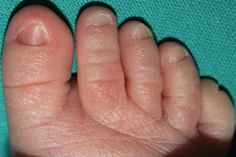
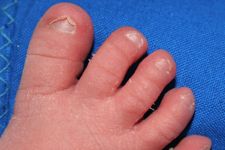
| Figure 1. Triangular shaped nail of the hallux in a 3-day-old baby. Figure 2. Apparent hypertrophy of the nail folds in a 2-day-old boy. | |
In 347 of the 541 cases (64.1%) the hallux nail was triangular shaped (Figure 1) or rarely trapezoidal, the shorter base being distal; in 194 cases it was rectangular shaped. With regard to gestational age, 68 of the 94 (72.3%) premature babies had triangular hallux nails compared with 279 of the 447 full term newborns (62.5%): the difference was not statistically significant (OR=1.57; chi-square=3.33; p=0.06; IC=0.95-2.68). The same alterations were sometimes also observed in other toenails but never in fingernails. The cases with a triangular shaped hallux nail also showed apparent hypertrophy of the lateral and/or distal fold of the periungual skin (Figure 2). At no time was inflammation of the periungual tissue observed.
 |  |
| Figure 3a | Figure 3b |
|---|---|
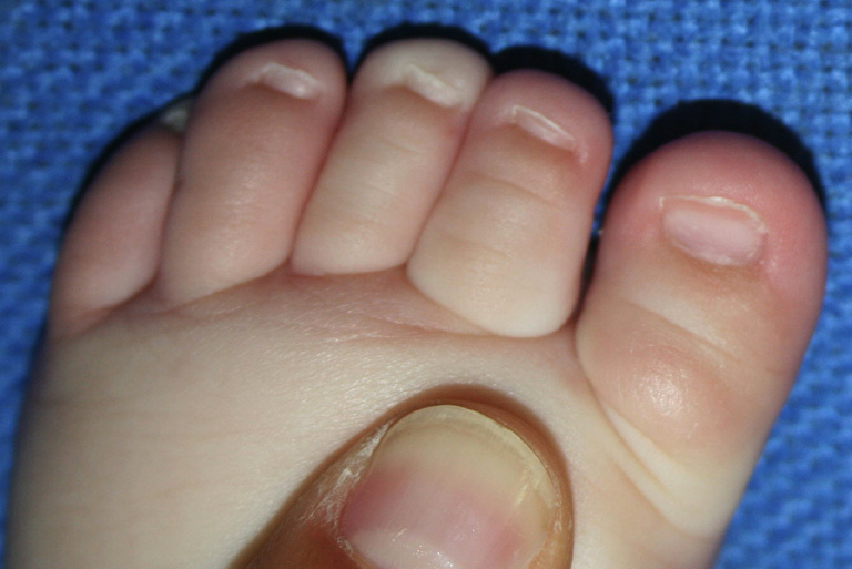
| Figures 3a and 3b. (a) Triangular shape of the hallux nail at the age of three days (a), turning into rectangular shape after 6 weeks (b) | |
In the subsequent control examinations, the triangular and trapezoidal hallux nails progressively became rectangular shaped (Figures 3a and 3b) in 45-90 days (average two months).
Discussion
The condition we called triangular nail of the hallux had been previously reported in the literature as congenital hypertrophy of the lateral fold of the hallux [2]. This condition can possibly be confused with “congenital ingrown toenail” [5, 6]. In the case of triangular nail of the hallux, at no time was inflammation of the periungual tissue observed and the condition spontaneously regressed in all cases examined in a few weeks. This ruled out the hypothesis of congenital ingrown toenail and supported the diagnosis of a physiological transitory alteration of the toenail. However, as we re-examined only about ten percent of the newborns with triangular nail of the hallux at birth, we cannot exclude that it can persist longer than a few weeks in some cases.
With regard to the previous name of congenital hypertrophy of the lateral fold of the hallux, the question is whether the hypertrophy is primary or secondary. Nails are an appendage of the epidermis. They begin to develop at 8-9 weeks of fetal development [7, 8, 9] and have slow and progressive growth. Toenails grow slower (1 to 1.5 mm per month) than fingernails (3 mm per month) [10] and growth of the nail of the big toe is minimal [11]. The slower growth of the toenails explains why at birth the triangular shape is only seen in the toenails and not in the fingernails. The triangular shape of the nail is probably related to the more rapid growth of the nail matrix in its central portion or to an asynchronous onset of growth of the nail matrix, starting in the center and then proceeding laterally. It is more evident at birth in the toenails, particularly in the nail of the “big toe,” because of its slower growth.
The very frequent condition we called triangular nail of the hallux was different from “great toenail dystrophy” [12], renamed “congenital malalignment of the big toenail” by Baran [13]. Our condition did not have lateral deviation of the long axis of nail growth, discoloration, thickening, or any other alterations of the nail. Moreover, it regressed in all the thirty-six re-examined newborns more rapidly than the condition of great toenail dystrophy [14].
In conclusion, the triangular shape of the big toenail is likely an expression of the slower and thus incomplete development of this nail at birth. It is also likely that the apparent hypertrophy of the distal and lateral folds of the nails of the big toe is only a secondary phenomenon and derives from the same causes as those responsible for the triangular shape.
References
1. Mari F, Politi L, Bertol E. Nails of newborns in monitoring drug exposure during pregnancy. Forensic Sci Int. 2008 Aug; 179(2-3):176-80, 2008. [PubMed]2. Piraccini BM, Parente GL, Varotti E, Tosti A. Congenital hypertrophy of the lateral nail folds of the hallux: clinical features and follow-up of seven cases. Pediatr Dermatol. 2000 Sep-Oct; 17(5):348-51. [PubMed]
3. Yinnon AM, Matalon A. Koilonychia of the toenails in children. Int J Dermatol. 1988 Dec; 27(10):685-7. [PubMed]
4. Turano AF. Transverse nail ridging in early infancy. Pediatrics. 1968 May; 41(5):996-7. [PubMed]
5. Honig PJ, Spitzer A, Bernstein R, Leyden JJ. Congenital ingrown toenails. Clinical significance. Clin Pediatr. 1982 Jul; 21(7):424-6. [PubMed]
6. Viseux V, Plantin P. L’ongle du nuveau-né et du nourrisson. Ann Dermatol Venerereol. 2003 Jan; 130(1):74-8. [PubMed]
7. Zaias N. Embryology of the human nail. Arch Dermatol. 1963 Jan; 87:37-53. [PubMed]
8. Hashimoto K, Gross BG, Nelson R, Lever WF. The ultrastructure of the skin of human embryos. III. The formation of the nail in the 16-18 weeks old embryos. J Invest Dermatol. 1966 Sep; 47(3):205-17. [PubMed]
9. Holbrook KA. Embryogenesis of the skin. In: Textbook of Pediatric Dermatology, Harper J, Oranje A, Prose N. eds, 2nd ed, Blackwell Publishing, 2006, pp. 3-41.
10. Runne U, Orfanos CE. The human nail: structure, growth and pathological changes. Curr Probl Dermatol. 1981; 9:102-49. [PubMed]
11. Pfister R, Heneka J. Wachstum und Gestaultung der Zehennagel bei Gesunden. Arch Klin Exp Dermatol. 1965 Sep 29; 223(3):263-74. [PubMed]
12. Samman PD Great toenail dystrophy. Clin Exp Dermatol 1978 Mar; 3(1):81-2. [PubMed]
13. Baran R Significance and management of congenital malalignment of the big toenail. Cutis 1996 Aug; 58(2):181-4. [PubMed]
14. Dawson TAJ Great toe-nail dystrophy. Br J Dermatol 1989 Jan; 120(1):139-40. [PubMed]
© 2010 Dermatology Online Journal