Phytophotodermatitis due to
Published Web Location
https://doi.org/10.5070/D3046507z8Main Content
Phytophotodermatitis due to Ficus carica
Muhterem Polat MD, Pınar Öztaş MD Asc Prof, Meltem Cik Dikilitaş MD, Nuran Allı MD Asc Prof
Dermatology Online Journal 14 (12): 9
Ankara Numune Education and Research Hospital, 1st Dermatology Department, Ankara, Turkey. drmuhterempolat@mynet.comAbstract
The genus Ficus belongs to the Moraceae (the mulberry family). Figs can cause irritant or phototoxic reactions. Phytophotodermatitis is a common cutaneous phototoxic reaction. Contact with plant-derived phototoxic substances (furocoumarins or psoralens) followed by sunlight exposure produces the clinical lesions. Here, we present a case of phytophotodermatitis after contact with fig fruits and leaves. The vesicular dermatitis was primarily located in areas of vitiligo.
Introduction
Figs can cause irritant or phototoxic reactions. These reactions occur in those who cultivate, gather, pack, or consume figs [1]. Furocoumarins have been detected in parts of Ficus carica. Furocoumarins are more plentiful in leaf sap than in shoot sap [2, 3]. Furocoumarins have been identified in the shoot sap and fruit , but no photoactive coumarins were detected in the seeds, peels, or fruit of either ripe or unripe figs [3, 4]. Phytophotodermatitis is a well-known entity that is caused by sequential exposure to certain species of plants containing furocoumarins and then to sunlight [2, 3, 4]. There are case reports of phytophotodermatitis caused by fig leaf decoction [5, 6]. Here, we present a case of phytophotodermatitis caused by exposure to Ficus carica.
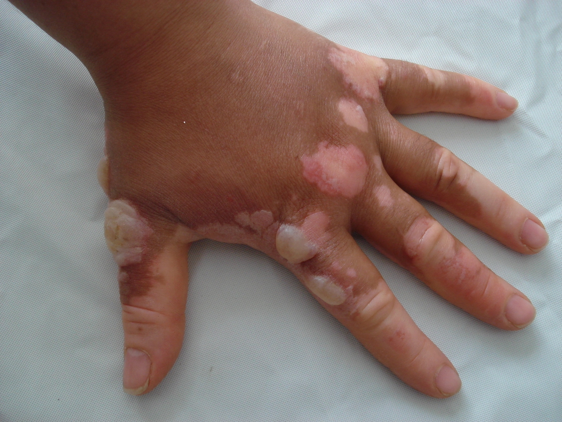 | 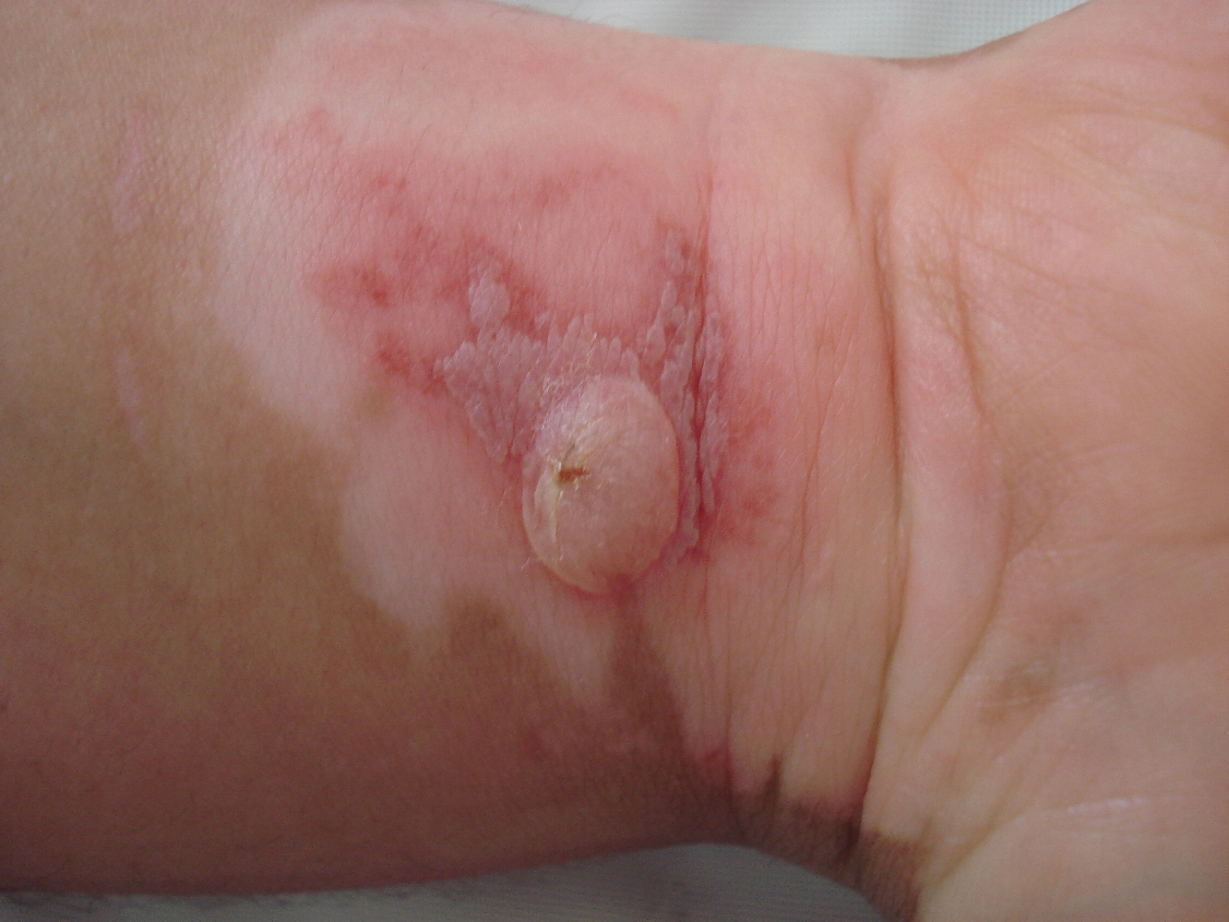 |
| Figure 1 | Figure 2 |
|---|---|
| Figure 1. Erythema, edema, bullae, and vitiligo patches are seen on the dorsum of hand Figure 2. Erythema and bullae on the anterior aspect of wrist | |
Case report
A 34-year-old woman working as a cook in a catering service presented to our outpatient clinic with erythema, edema, and large bullae on the dorsum of her hands. She reported that two days before presentation, she sliced many fig fruits, separated many leaves from the fruits, and exposed her hands to direct sunlight that day and the following day. On the second day, a burning sensation, itch, pain, erythema, edema, and subsequently bullae appeared on both hands. The patient presented on the morning of the third day with erythema, edema, and large bullae on the dorsum of her hands. Dermatological examination also revealed the presenc of vitiligo on the hands and trunk. Bullae were localized to the vitiligo areas. Cultures obtained from the bullae fluid were negative. Treatment included aspiration of the bullae, application of topical antibacterial creams and wet dressings, and the administration of systemic antihistamines. The patient did not agree to patch testing. Her lesions began to recover after five to six days and resolved completely within ten days.
Discussion
Phytophotodermatitis is a common cutaneous phototoxic reaction. Contact with plant-derived phototoxic substances (furocoumarins or psoralens) followed by sunlight exposure produces the clinical lesions. These phototoxic substances are found in various vegetable families.
The genus Ficus belongs to the Moraceae (mulberry) family. Ficus carica can grow among rocks, in woods, and in hot, dry soils [1]. Irritant dermatitis has been reported from contact with small hairs on the undersurface of Ficus carica leaves. The latex contains ficin, a proteolytic enzyme that causes pruritus, and acts as an irritant on inflamed skin. The milky latex found in the leaves and stems seems to contain the irritant and phototoxic chemicals [3, 4]. There are case reports of phytophotodermatitis caused by fig leaf decoction [5, 6]. Two studies have detected furocoumarins in parts of Ficus carica [2,3]. In both studies, fig leaves were examined. Examination revealed that psoralens (parent compounds in the furocoumarin family) and bergapten (5-methoxypsoralen) were present throughout the growing season. Furocoumarins usually are more plentiful in leaf sap than in shoot sap and concentrations are higher in spring and summer than in fall [2]. Zaynoun et al. [3] studied the presence of furocoumarins in the shoot sap and fruit and found no photoactive coumarins in the seeds, peels, or fruit of ripe or unripe figs. This seems to contradict a study conducted by Ippen [4] demonstrating clinical phytophotodermatitis following contact with fresh figs. Enzymes such as proteases, lipodiastases, and amylase, which have keratolytic effects, are also found in the fig latex and may enhance the phototoxic effects of furocoumarins [3].
Our patient had contact with both fruits and leaves of figs. She was probably sensitized with either the fruit or leaf or both of them and her clinical findings and course suggested the diagnosis of phytophotodermatitis. The localization of bullae to areas of dermatitis probably reflects the decreased protection from sunlight in these areas.
Phytophotodermatitis may occur as an occupational hazard anywhere in the world. It is prudent to advise the use of gloves and protective clothing while working with plants containing furocoumarins and to avoid simultaneous exposure to sunlight.
References
1. McGovern TW. Botanical Briefs: The Fig-Ficus carica L. Cutis. 2002; 69: 339-340. [PubMed]2. Innocenti G, Bettero A, Caporale G. Determination of the coumarinic constituents of Ficus carica leaves by HPLC. Farmaco (Sci). 1982; 37: 475-85. [PubMed]
3. Zaynoun ST, Aftimos BH, Abi Abi L, et al. Ficus carica: isolation and quantification of the photoactive components. Contact Derm. 1984; 11: 21-5. [PubMed]
4. Ippen H. Phototoxic reaction to figs. Hautarzt. 1982; 33: 337-9. [PubMed]
5. Ozdamar E, Ozbek S, Akın S. An unusual cause of burn injury: Fig leaf decoction used as a remedy for a dermatitis of unknown etiology. J Burn Care Rehabil. 2003; 24: 229-33. [PubMed]
6. Bollero D, Stella M, Rivolin A, Cassano P, Vanzetti M. Fig leaf tanning lotion and sunrelated burns: case reports. Burns. 2001; 27: 777-9. [PubMed]
© 2008 Dermatology Online Journal