|
| Figure 1 |
|---|
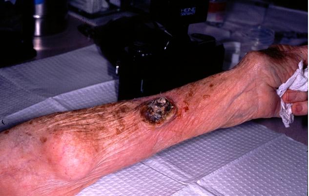
| Figure 1. Tumor on volar aspect of left forearm. |
An 82-year-old white woman presented with a raised, 3x4 cm, painless growth on her left forearm. The lesion had been present for approximately 2 months and was not associated with any symptoms. The patient had a history of a previous skin cancer on her lower extremity. Physical examination demonstrated a cone-shaped keratotic cutaneous horn as shown in Figure 1 & 2. Histopathologic findings are shown in Figure 3.
 |
 |
| Figure 2 | Figure 3 |
|---|---|
| Figure 2. Close-up view of the tumor surmouted by cutaneous horn. | |
| Figure 3. Histology of the lesion. | |
The biopsy specimen revealed a neoplastic proliferation of atypical keratinocytes extending into the dermis, with large, hyperchromatic and pleomorphic nuclei.
The term cutaneous horn, or cornu cutaneum, refers to a well-defined cone-shaped lesion with hyperkeratotic features.[
1,2] In general, cutaneous horns (whether associated with benign or malignant lesions) are found most frequently on exposed skin. Various types of associated lesions can be found at the base of a cutaneous horn, including squamous cell carcinoma, actinic keratosis, keratoacanthoma, Bowen's disease, seborrheic keratosis, basal cell carcinoma, hemangioma, keratotic and micaceous pseudopapillomatous balanitis, Kaposi's sarcoma, sebaceous adenoma and Paget's disease of the female breast.[3,4] In all, approximately 20% of cutaneous horns are associated with malignancy.[5,6] In one study of 35 patients with 37 cutaneous horns, 21 (56%) were benign, and 14 (44%) were malignant. The study also demonstrated that of the 20 patients with cutaneous horns in association with benign lesions, one had a history of a basal cell epithelioma, another had a history of Bowen's disease, and a third patient had a previous squamous cell carcinoma. Thus 15% of these patients had a history of associated skin malignancy.[3] Of the 15 patients with cutaneous horns in association with malignant or pre-malignant lesions, three patients had a history of basal cell-epitheliomas and two patients had previous squamous cell carcinomas. Therefore, 33% of these patients had a history of previous skin malignancies. These data suggest that a cutaneous horn associated with a malignant or pre-malignant base is more common in patients with a history of other malignant or pre-malignant lesions.[3]© 2000 Dermatology OnLine Journal