|
| Figure 1 |
|---|
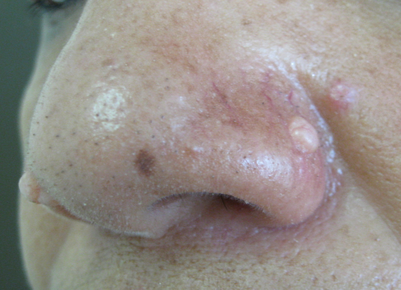
| Figure 1. Two papules are located at both free-edges of the nose, well demarcated, lobulated, showing a slightly pink color, with visible telangiectasias on their surface. |
A healthy, 46-year-old female presented with a 7-year history of two lesions, which had been undergoing steady growth, located symmetrically at both sides of the nose.. On physical examination, there were two papules, measuring 0.3 and 0.4 cm in diameter. They were well demarcated, smooth-surfaced, and lobulated; they showed a slightly pink color, with visible telangiectasias on their surface (Figure 1).
 |
 |
| Figure 2 | Figure 3 |
|---|---|
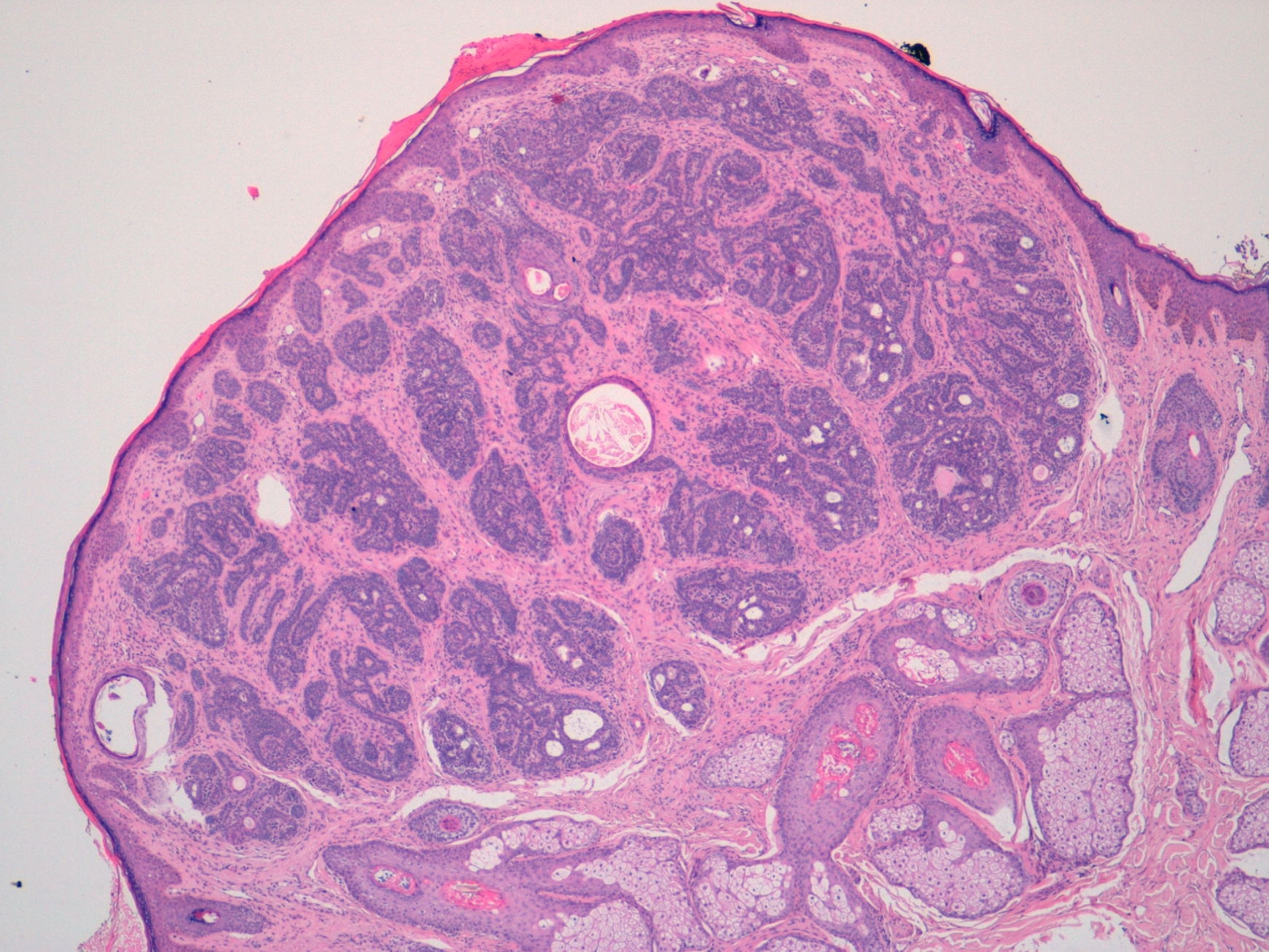
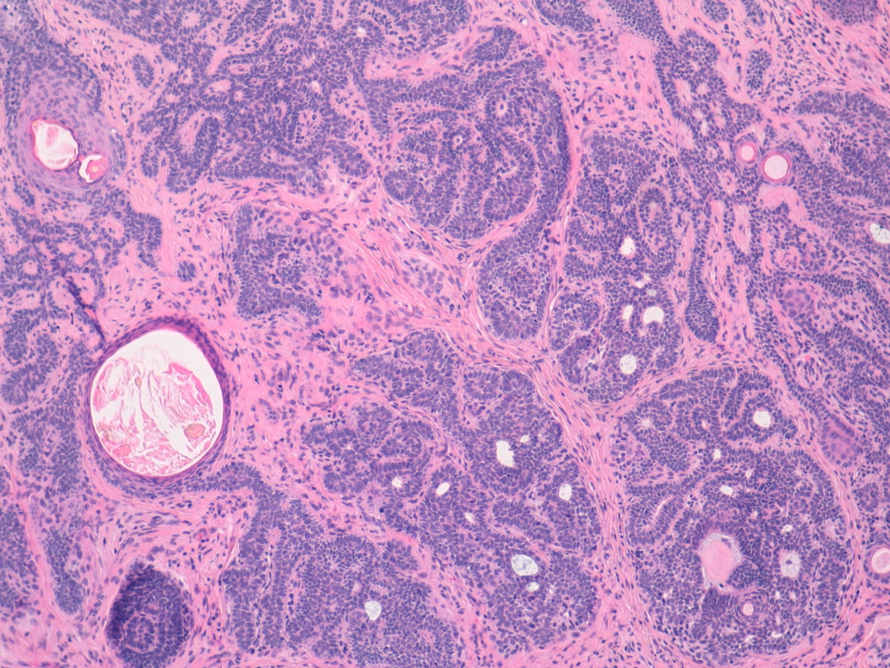
| Figure 2. Histological section of the tumor showing a well-demarcated, symmetrical tumor (H&E, x4) Figure 3. Detail of the lesion showing lobules of small, irregular basaloid cells, surrounded by a fibrous cellular stroma with a moderate lymphocytic infiltrate (H&E, x10). |
|
Histological examination revealed a well-demarcated tumor composed of lobules of small, irregular basaloid cells showing follicular differentiation, surrounded by a fibrous cellular stroma with a moderate lymphocytic infiltrate (Figures 2 and 3).
Trichoblastomas are rare, benign tumors of the hair germ composed of follicular germinative cells. These are deeply or superficially situated dermal nodules, found on the head and neck. They usually present as solitary skin-colored nodules that are variable in size (usually 1 or 2 centimeters). They can also present as pigmented papules, usually arising from a Jadassohn sebaceous nevus [1].
Nests of basophilic basaloid cells with a lobular architecture and prominent induction of stroma are seen in the dermis and/or subcutaneous tissue. Focal evidence of follicular differentiation is seen, but this usually consists of less mature structures than those seen in trichoepithelioma. Mitotic figures are frequent. Usually, the tumor is not connected to the epidermis [2].
Clarification should be made with the entity termed trichoepithelioma. Ackerman et al have used trichoblastoma as a generic term for all neoplasms of the skin and subcutaneous fat that are composed mostly of follicular germinative cells. Trichoepitheliomas are included in this definition and thus considered as a histopathological variant of trichoblastomas. They have reported nodular, retiform, cribriform, racemiform, and columnar patterns of trichoblastoma. They share many architectural findings, such as the proliferation of basal germinative cells surrounded by a dense fibrous stroma [3]. As a distinctive finding, trichoblastoma shows no epidermal connection whereas trichoepitheliomas are dermal tumors with focal continuity with the epidermis in up to one-third of cases [4].
Trichoepitheliomas, when solitary, clinically present as skin-colored papules or small nodules located on the face, showing a special predilection for the nose. Multiple familial trichoepitheliomas present as small papules located mostly at the central part of the face. The papules may coalesce to form plaques. The onset of lesions is usually childhood. The presence of multiple trichoepitheliomas is seen in Brooke-Spiegler syndrome, an uncommon disease with a predisposition to develop cutaneous adnexal neoplasms such as cylindromas, trichoepitheliomas, spiradenomas, trichoblastomas, basal-cell carcinomas, follicular cysts, organoid nevi; there may be malignant transformation of pre-existing tumors in the affected individuals. Brooke-Spiegler syndrome is inherited in autosomal-dominant fashion [5]. The gene for multiple trichoepitheliomas has recently been mapped to a locus on chromosome 9p21 [6]. Trichoepitheliomas are benign lesions. The histopathology comprises elements of basal cell carcinoma and trichoepithelioma with a variable degree of follicular differentiation. However, the stroma induced in trichoepithelioma is distinctive and contains clefts, and there is absence of retraction artefact between tumor cells and the surrounding stroma.
This case shows an atypical presentation of trichoblastomas because they usually are not so well demarcated and when they are multiple, they tend to be small and clustered around the nose. However, histological and clinical findings allowed us to establish the appropriate diagnosis.
© 2011 Dermatology Online Journal