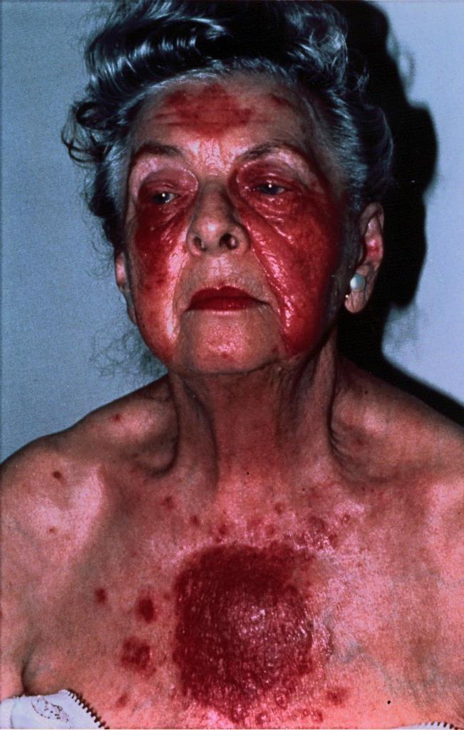
History 77-year old woman referred for evaluation of an erythematous, partially denuded eruption over her face and trunk of 15 months duration. The eruption began with an enlarging area of red- ness confined to her left cheek and spread despite treatment with antibiotics, topical and intra-lesional corticosteroids. She had no constitutional symptoms throughout the course of her illness. Laboratory eval- uations including skin cultures were negative. Three consecutive skin biopsies were interpreted as psoriasiform dermatitis.
Physical Examination She had well-demarcated ery- thematous and partially denuded plaques in a seborrheic distri- bution over her face, chest, and back. Several 2-4mm flaccid vesicles were present within the facial plaques.
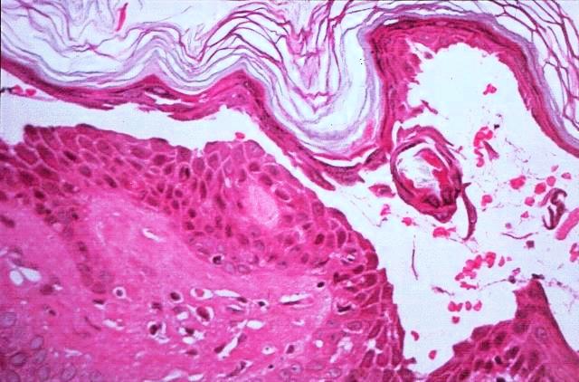
Histology A punch biopsy of involved facial skin revealed acantho- lysis within the granular layer layer forming a subcornea cleft, and a sparse perivascular lymphoplasmacytic infiltrate in the papillary dermis.
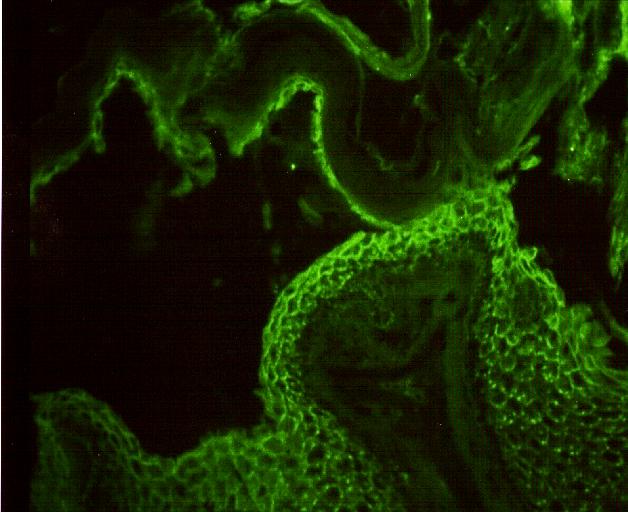
Immunofluorescence Direct immunofluorescence studies revealed IgG within intercellular spaces of the epidermis and C3 confined to those granular layer keratinocytes forming the floor of the cleft. Indirect immuno- fluorescence for pemphigus antibody was present at a titer of 1:40.