|
| Figure 1 |
|---|
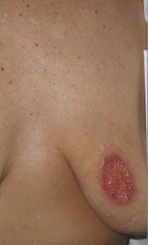
A 41-year-old female presented for evaluation of a painful ulcer on her left breast. The ulcer developed 6 months prior to presentation and had been draining purulent material. The patient was treated with numerous courses of antibiotics without any response. Serial cultures from the ulcer have all been negative. At the time of presentation, patient complained of soft tissue swelling at the root of the nose, occasional epistaxis, generalized malaise and a 15-pound weight loss over 5 months.
Physical examination of her left breast revealed a 5 by 2.5 cm oval irregular ulceration with violaceous undermined borders (Fig. 1).
 |
 |
| Figure 2 | Figure 3 |
|---|
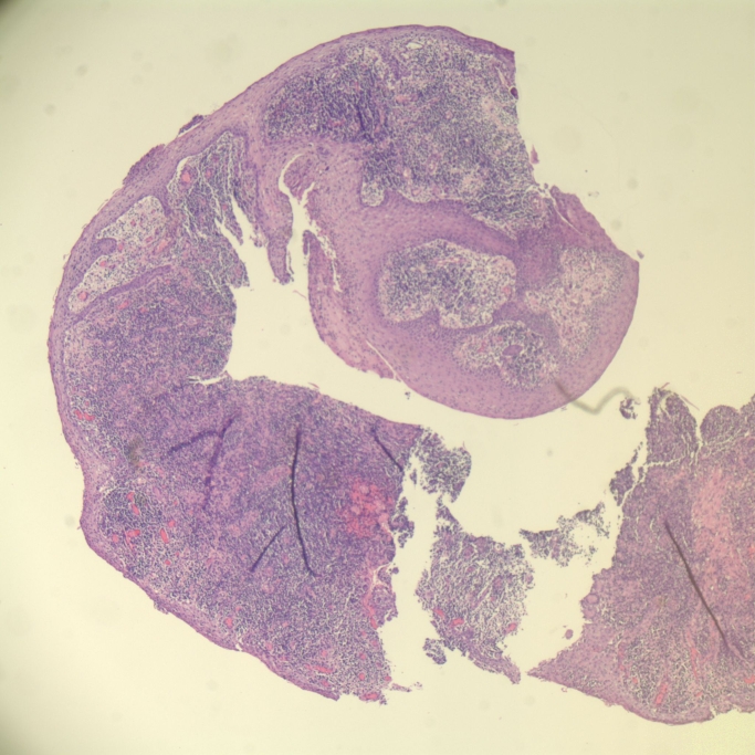
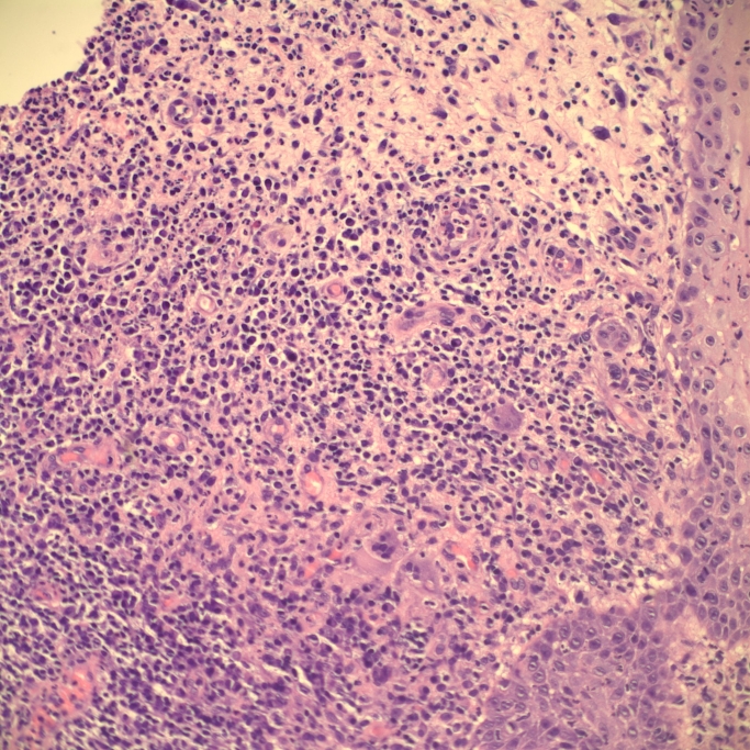
Biopsy of the lesion showed a focally acanthotic and ulcerated epidermis with intense granulomatous and mixed dermal infiltrate consisting of lymphocytes, histiocytes, plasma cells, neutrophils and scattered multinucleate giant cells (Fig. 2). Focal vascular necrosis with extravasation of erythrocytes and vascular proliferation was also observed (Fig. 3). Special stains for bacteria, fungi, and mycobacteria were all negative.
Further workup revealed an elevated cytoplasmic anti-neutrophil cytoplasmic antibody (cANCA) with anti-proteinase antibody titer grater than 19 units/ml, erythrocyte sedimentation rate (ESR) of 104 mm/hr, Hemoglobin 10.3 with MCV of 77.3, platelets of 684,000 unremarkable blood urea nitrogen (BUN), creatinine and urinalysis. Subsequent nasal endoscopy showed a total perforation of the nasal septum. Chest x-ray was negative for pulmonary pathology.
A diagnosis of Wegener's granulomatosis (WG) was made and patient was started on prednisone and oral methotrexate. After 2 months of therapy the ulcer has completely resolved. Followup ESR, platelets, hemoglobin and cANCA also returned to baseline.
Cutaneous involvement in WG is common and occurs in 40-50 percent of patients [1]. Clinically, purpura is the most common dermatologic manifestation of WG; in the majority of cases this represents necrotizing vasculitis [2, 3]. Pyoderma gangrenosum (PG)-like ulcerations are less common; they show granulomatous inflammation which may or may not be associated with underlying vasculitis [1, 3].
PG-like lesions usually either precede systemic symptoms of WG or occur very early in the disease [1]. Majority of Wegener's PG-like lesions have been described in young men that developed multiple lesions usually on the head and neck [4].
Unlike most of PG-like manifestations of WG that are present in men with diffuse cutaneous disease, our female patient had a single lesion on the breast and in that respect does not fit into typical PG-like cutaneous WG noted by other authors. Overall, breast involvement in WG is exceedingly rare and in most of the reports has been described as breast masses or nodules with occasional focal ulceration rather than PG-like lesions [5]. The diagnosis in our patient was made after thorough evaluation of all pieces of data including clinical history, biopsy of the ulcer, radiological findings, and serological evaluation. This case further supports aggressive diagnostic approaches to PG-like ulcers, especially in the absence of response to therapy.
© 2007 Dermatology Online Journal