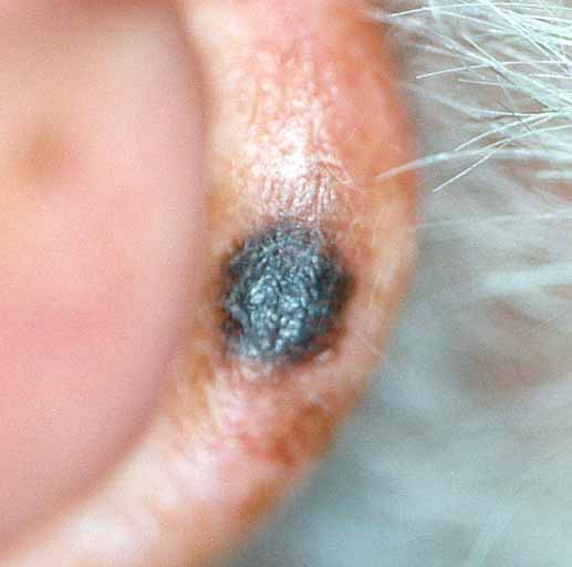
Figure 7
A pigmented 6 mm papule is present on the left ear helix. The lesion is symmetrical, homogenous in color, and has a smooth border.
![]()
A 55-year-old man presented with a two-year history of a pigmented lesion of the left ear helix. Examination showed a 5.5-mm, symmetrical, dark brown, homogeneous macule with a smooth border (Figure 7).
|
Figure 7 A pigmented 6 mm papule is present on the left ear helix. The lesion is symmetrical, homogenous in color, and has a smooth border. |
|
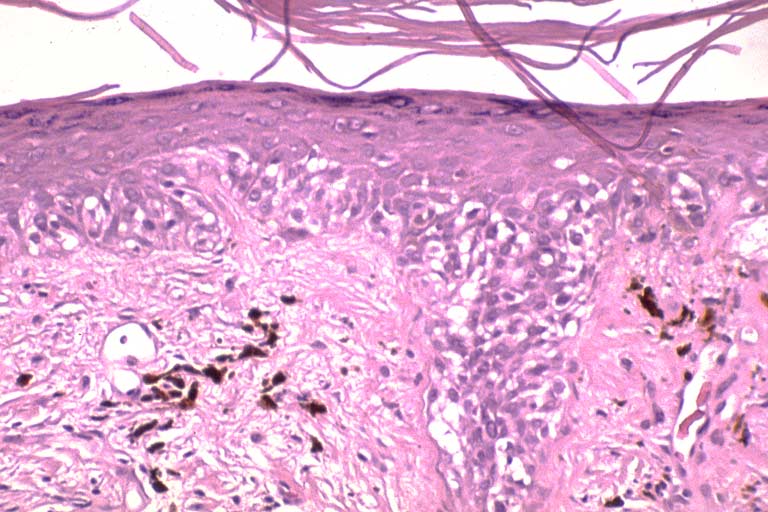
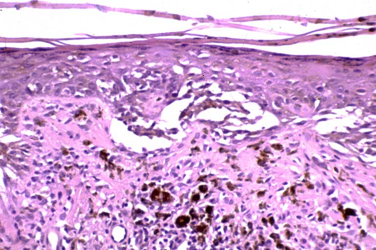
It was thought to be a seborrheic keratosis. One month later, the patient returned noting that the lesion had slowly grown; punch excision was performed. Histopathologic investigation showed a proliferation of melanocytes at the dermal-epidermal junction forming irregular nests. The melanocytes showed variation in nuclear shape, size, and chromatin pattern and were present at higher levels within the epidermis, and followed the dermal-epidermal junction into hair follicles (Figures 8a, 8b).
|
Figure 8a
A medium-power view showing in situ melanoma presenting as atypical lentiginous melanocytic hyperplasia. |
|
|
Figure 8b
A medium-power view af another area showing pagetoid spread of atypical melanocytes. |
|
No invasive growth was present. The diagnosis of malignant melanoma in situ was made.
© 1999 Dermatology Online Journal