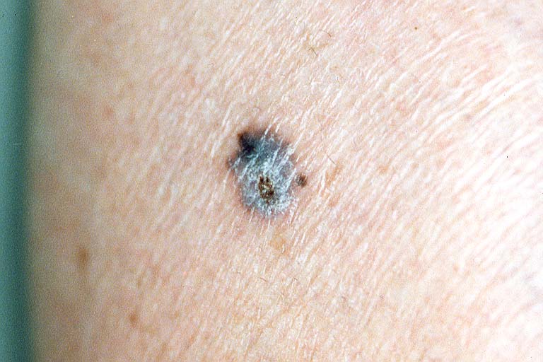
A uniformly pigmented, blue-black, 6 mm papule present on the left lower leg. There is scaling, focal crusting and a smooth border.
![]()
A 51-year-old female presented with a 10-year history of a pigmented lesion on the left lower leg. Examination showed a symmetrical, pigmented lesion 6 mm in diameter, with gray coloration, scaling surface, focal crusting, rim of erythema and a smooth border (Figure 5).
|
Figure 5
A uniformly pigmented, blue-black, 6 mm papule present on the left lower leg. There is scaling, focal crusting and a smooth border. |
|
None of the patient's other lesions appeared crusted or inflamed. Clinically, it was thought to be an irritated melanocytic nevus; however the history of persistent crusting led to a punch excision.
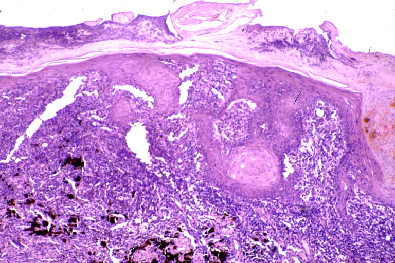
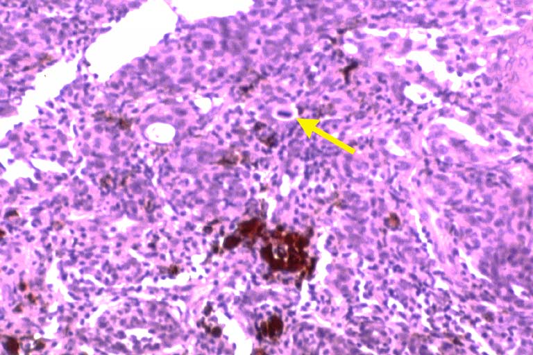
Histopathologic investigation demonstrated irregular proliferation of atypical melanocytes at the dermal-epidermal junction with invasion into the dermis to a depth of 0.65 mm (Breslow), Clark's Level III (Figures 6a, 6b).
|
Figure 6a
A scanning magnification view intended to show, in the left one half, an intradermal sheet of atypical melanocytes interspersed with melanophages. |
|
|
Figure 6b
A close-up view highlighting a mitotic figure (arrow) in the center of the intradermal sheet of atypical melanocytes. |
|
The diagnosis of malignant melanoma was made.
© 1999 Dermatology Online Journal