A pigmented papule measuring 5 mm in diameter is present on the right posterior leg. It is modestly asymmetric but fails to show irregular border or variegation of color.

![]()
A 75-year-old woman presented with a one-year history of a pigmented, 5-mm lesion on the right posterior leg. The lesion had recently enlarged and darkened. Examination showed a nearly symmetrical homogenous, dark-brown, papule with a smooth border (Figure 3).
|
Figure 3
A pigmented papule measuring 5 mm in diameter is present on the right posterior leg. It is modestly asymmetric but fails to show irregular border or variegation of color. |
|
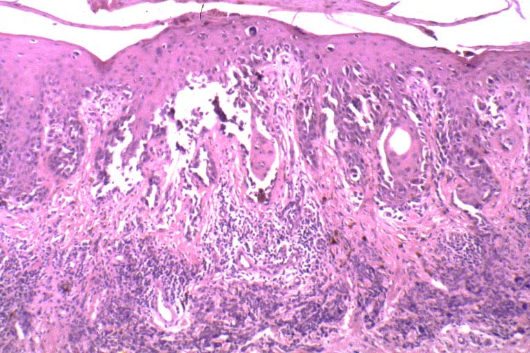
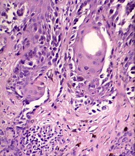
Clinically, the first diagnosis considered was melanocytic nevus; however, the lesion appeared different than the patient's other moles, and her history of recent onset and progressive increase in size with darkening of color led to a punch excision. Histopathologic investigation showed proliferation of atypical melanocytes forming irregular nests at the dermal-epidermal junction. Some melanocytes were found at elevated levels within the epidermis (Figures 4a, 4b, 4c).
|
Figure 4a
A scanning magnification view showing confluent proliferation of cytologically atypical melanocytes along the basal cell layer with a mild degree of pagetoid spread. The dermis shows an intradermal nevus. |
|
|
Figure 4b
A close-up view highlighting the atypical lentiginous melanocytic hyperplasia. |
|
|
Figure 4c
A close-up view of another area showing pagetoid spread. |

|
No invasive malignant melanoma was present. The diagnosis of malignant melanoma in situ was made.
© 1999 Dermatology Online Journal