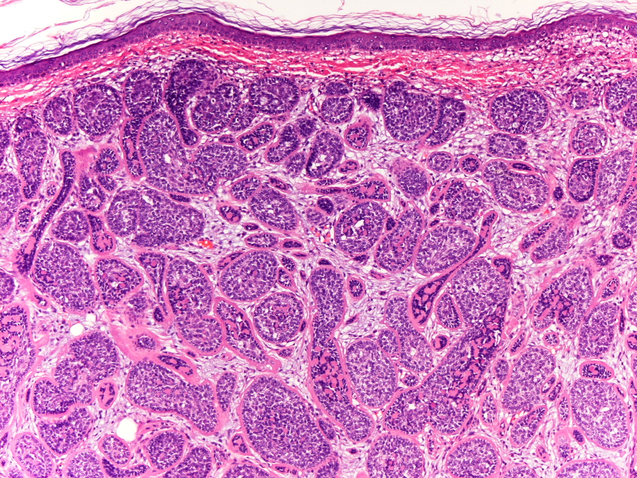
Figure 2. Dermal proliferation of basophilic lobules which molded together in a jigsaw puzzle configuration (H&E, x100)
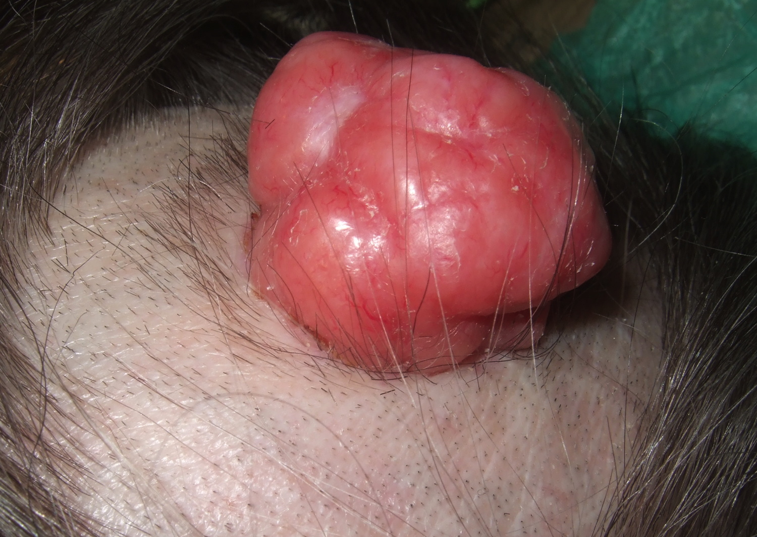
A 77-year-old healthy woman presented with an isolated exophytic lesion of the scalp for 4 years, which had grown progressively. On exam, there was a 4 cm, firm erythematous and lobulated tumor with telangiectasias on the scalp (Figure 1). The rest of her cutaneous exam was normal. No lymphadenopathy was appreciated. A solitary cylindroma was diagnosed by biopsy.
|
|
| Figure 1 | Figure 2 |
|---|---|
| Figure 1. Exophytic lesion of the scalp Figure 2. Dermal proliferation of basophilic lobules which molded together in a jigsaw puzzle configuration (H&E, x100) |
|
 |
| Figure 3 |
|---|
| Figure 3. Higher magnification reveals at the periphery, there was a thick eosinophilic PAS+ basement membrane material (type IV collagen) (H&E, x200). |
A 77-year-old healthy woman presented with an isolated exophytic lesion of the scalp for 4 years which had grown progressively. On exam, there was a 4 cm erythematous and lobulated tumor with telangiectasias and hard consistency on the scalp (Figure 1). The rest of her cutaneous exam was normal. No lymphadenopathy was appreciated. Histologic sections are shown in Figures 2 and 3.
Histological sections of skin showed a dermal proliferation of basophilic lobules which molded together in a jigsaw puzzle configuration (Figure 2). At the periphery, there was a thick eosinophilic PAS+ basement membrane material (type IV collagen). The nests were also punctuated by small round collections of this material with similar staining qualities. Cells with prominent vesicular nuclei were visible in the center of the molded lobules. Nodules formed by clear cells, basophils, and deposits of basement membrane between basophils cells corresponding to spiroadenoma were also seen (Figure 3).
The erythematous nodule was treated with surgical excision without any evidence of recurrence after seven months of follow-up.
Dermal cylindromas are usually benign neoplasms of the eccrine sweat glands. The tumor usually occurs in the sixth decade of life [1]. There is no sex predilection. It presents most commonly on the head, neck, or scalp as slowly growing, pink to purple, solitary or multiple, smooth surfaced nodules, which can rarely grow and coalesce to produce the characteristic turban-like mass (turban tumor) [2]. The Brooke-Spiegler syndrome is an uncommon autosomal dominant disorder characterized by a high affinity to form multiple cylindromas, spiradenomas, and trichoepitheliomas that may rarely be associated with identical lesions in salivary glands, breast, or lung. The gene implicated in BSS, the CYLD gene, a tumor suppressor gene, is located on chromosome 16q [3]. In rare instances, malignant transformation affects multiple cylindromas more often than solitary cylindromas. The clinical signs of malignant transformation are ulceration, rapid growth, bleeding, and blue to pink coloration of the nodules [4].
Histological characteristics of cylindrocarcinoma show ill-defined areas of necrosis, nuclear pleomorphism, and a high mitotic index.
The differential diagnoses of tumors on the scalp may include benign growths, such as trichilemmal cyst, which is composed of squamous epithelium without a granular layer with abrupt homogeneous keratinization. In addition, malignant tumors, such as metastatic carcinomas may spread to the skin via blood or lymphatics. The most common sources are breast, colon, and bronchial carcinomas, as well as metastatic melanoma. In skin metastases, the epidermis is usually uninvolved and biopsy shows a nodular proliferation of malignant cells [5]. Squamous cell carcinoma is a malignant tumor characterized by irregular masses of atypical epidermal cells with infiltration of the dermis. Clinically they may reveal scaling, ulceration, crusting, or a cutaneous horn. Merkel cell carcinoma is a diffuse or trabecular basophilic tumor with medium-sized tumor cells with round somewhat dense vesicular nuclei and little cytoplasm. Merkel cell carcinoma commonly shows apoptotic cells and mitoses. It expresses cytokeratins, especially CK20, with a perinuclear punctate pattern, and neurofilament, synaptophysin, chromogranin, and CD56 [6]. Proliferating tricholemmal tumor is a benign tumor originating from the outer root sheath of a hair follicle. It is usually a solitary lesion and most commonly occurs in elderly women. Although considered biologically benign, it may be locally aggressive. In rare instances, malignant transformation has been reported, evidenced by regional or distant metastases [7].
© 2010 Dermatology Online Journal