Photoessay: The Skin and Diabetes Mellitus
by A Huntley
Dermatology Online Journal, December 1995
Volume 1, Number 2
Diabetes and Thick Skin
finger pebbles
additional images of pebbles
additional histology of pebbles
diabetic scleredema
diabetic hand syndrome
pebbles - acanthosis nigricans and diabetes
Shortly after a prospective evaluation of the skin findings of diabetes mellitus was initiated, a patient presented with
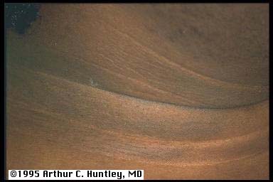
diabetes and acanthosis nigricans. He had typical velvety skin involving his neck and axillary region, and his acanthosis
nigricans was so exuberant that it involved the dorsum of his hands.


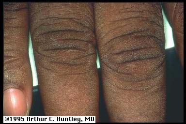
Figs 1,2. Anterolateral neck region and dorsum of right hand of a diabetic patient with acanthosis nigricans. Note the involvement
of the neck region recognized by the darker velvety skin. The dorsum of the fingers are similarly involved with velvety skin.
Involvement of the dorsum of the hands and fingers is known to occur in patients with advanced acanthosis nigricans. The velvety
appearance of the skin is similar to that seen in other involved skin areas where it is associated with increased epidermal
thickness.
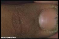
Fig 3. This close-up view of the dorsum of the finger from the above patient demonstrates on higher magnification that velvety
appearance consists of multiple closely spaced micropapules.
pebbles - thick skin of diabetes mellitus
The next few diabetic patients in the series did not have acanthosis nigricans (no neck or axillary involvement) but they
did have similar but less exaggerated changes on the dorsum of the fingers. This observation led to inclusion of this skin
change into the prospective evaluation.
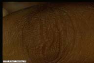
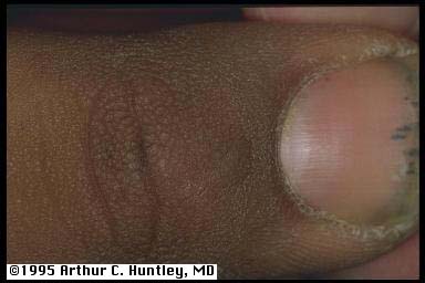
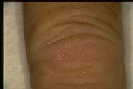
Fig 4. Dorsal finger skin of diabetic patient who does not have acanthosis nigricans. This diabetic patient was the next one
examined in the series. Although he does not have acanthosis nigricans, he does have similar discrete micropapules located
on the knuckle area of this finger..
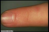
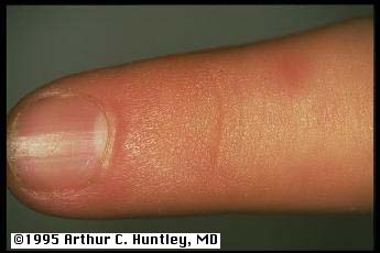
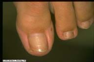
Fig 5. Proximal nail fold skin of a diabetic patient who demonstrates micropapules or "pebbles" in the absence of acanthosis
nigricans. Note that the micropapules in this patient are far less exaggerated than those which occur in acanthosis nigricans.
Finger pebbles were a common finding among the studies diabetic patients and it was postulated that they represent a physical
sign of thickened epidermis, similar to acanthosis nigricans.
finger pebbles - histopathology
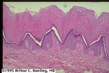
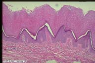 Fig 6. Histology sections of pebbled knuckle skin from this patient with diabetes demonstrates thickening of the epidermis
and papillary dermis, with coarsening of the papillary dermal collagen. The papules seen on histology correspond in size to
the clinical lesions..
Fig 6. Histology sections of pebbled knuckle skin from this patient with diabetes demonstrates thickening of the epidermis
and papillary dermis, with coarsening of the papillary dermal collagen. The papules seen on histology correspond in size to
the clinical lesions..
The histology appeared to confirm the presence of thickened epidermis, but also indicated some involvement of the papillary
dermis. Collagen fibers of the papillary dermis are broad similar to that seen with rubbed skin, only in these patients there
is no history of rubbing.
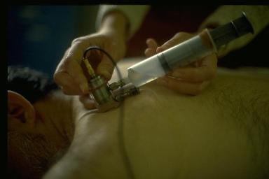
We then used ultrasound to measure skin thickness on diabetic subjects and found about half had significant increase in hand
skin thickness over that expected for age, but that the thickness did not correlate with duration of diabetes.


Figs 7,8. The ultrasound transducer is placed on the skin with a water interface as demonstrated here. The dermal thickness
is represented by the high peaked area, and is given as a digital readout.
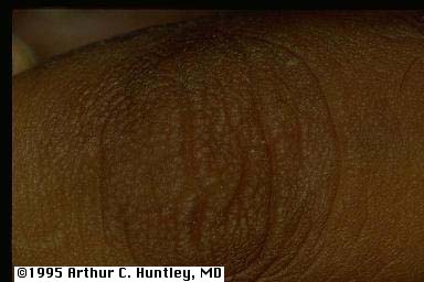
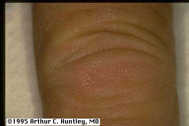
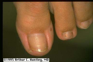
 Figs 9,10. Distal inter-phalangeal joint and dorsum of the distal aspect of the left foot in two diabetic patients demonstrating
pebbling. Clinically thickened skin is common on the dorsum of the hand (especially over the knuckles) and sometimes even
on the dorsum of the toes. Physical evidence of thickened skin is present in about half the patients with diabetes mellitus.
Figs 9,10. Distal inter-phalangeal joint and dorsum of the distal aspect of the left foot in two diabetic patients demonstrating
pebbling. Clinically thickened skin is common on the dorsum of the hand (especially over the knuckles) and sometimes even
on the dorsum of the toes. Physical evidence of thickened skin is present in about half the patients with diabetes mellitus.
In summary, pebbled skin over the dorsum of the knuckles and periungual area is a common finding in patients with diabetes
mellitus. The histologic sections indicate that this is associated with increased thickness of the epidermis and papillary
dermis. An ultrasound study also supports the concept that diabetics tend to have thicker skin in this area.
additional images of pepples
additional histology of pebbles
All contents copyright (C), 1995.
Dermatology Online Journal
University of California Davis