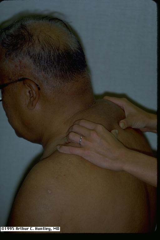
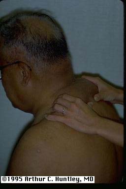
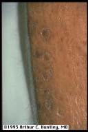
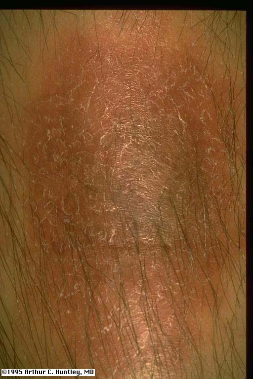
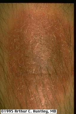
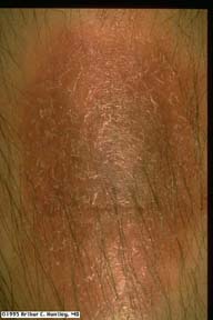
acanthosis nigricans in patient with diabetes mellitus fig1: acanthosis nigricans of the neck
L M S
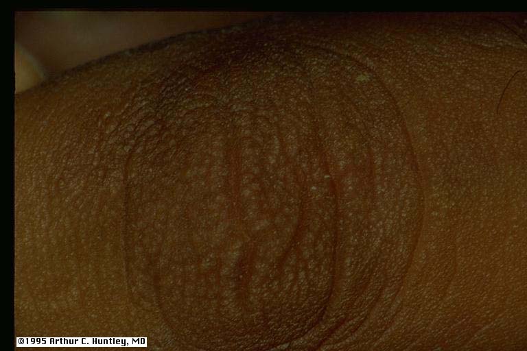
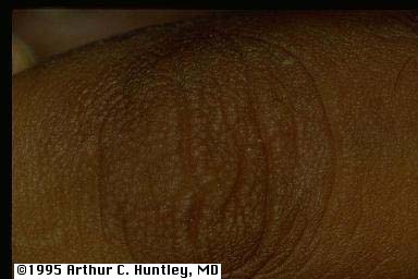
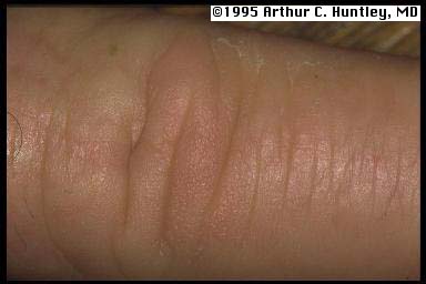
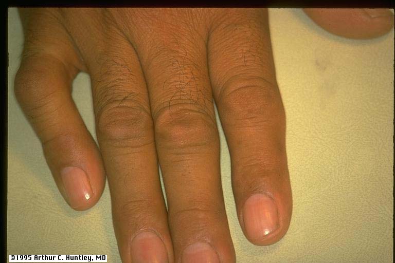
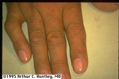
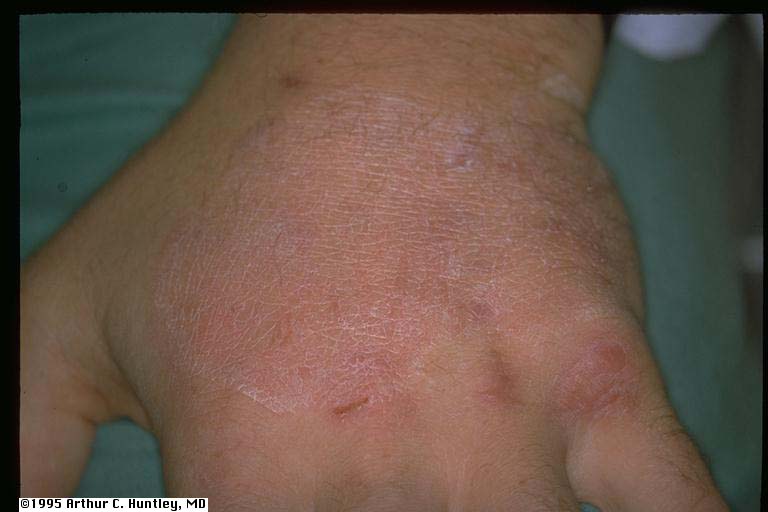
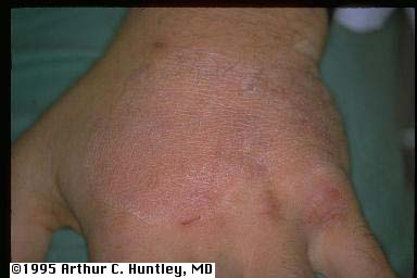
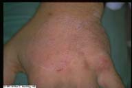
fig2: acanthosis nigricans of the dorsum of the hand
L M S
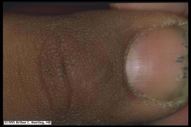
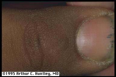
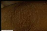
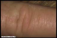
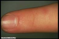
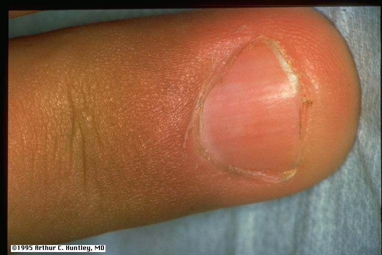
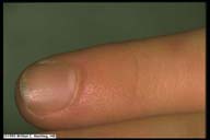
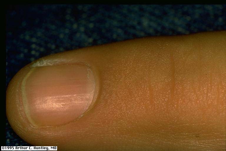
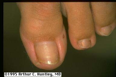
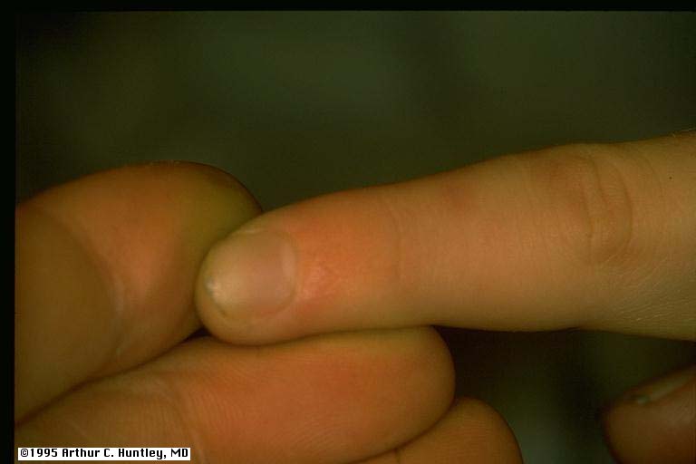
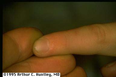
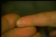
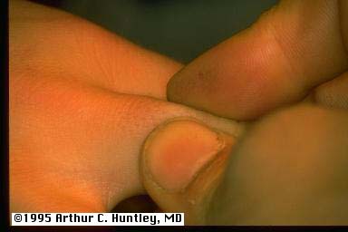
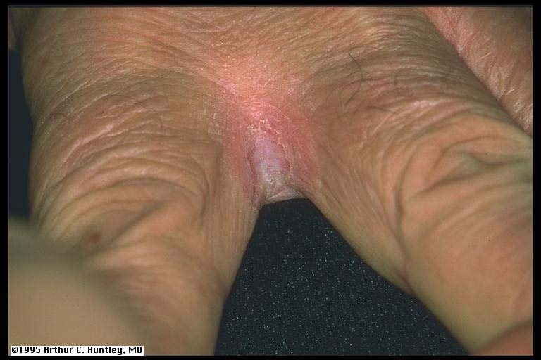
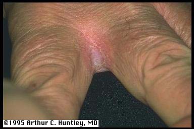
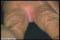
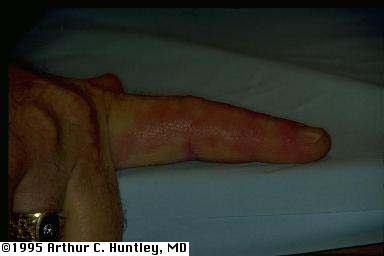
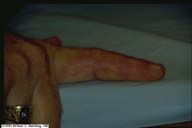
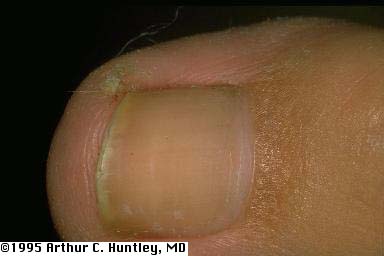
finger involvement of acanthosis nigricans - close-up
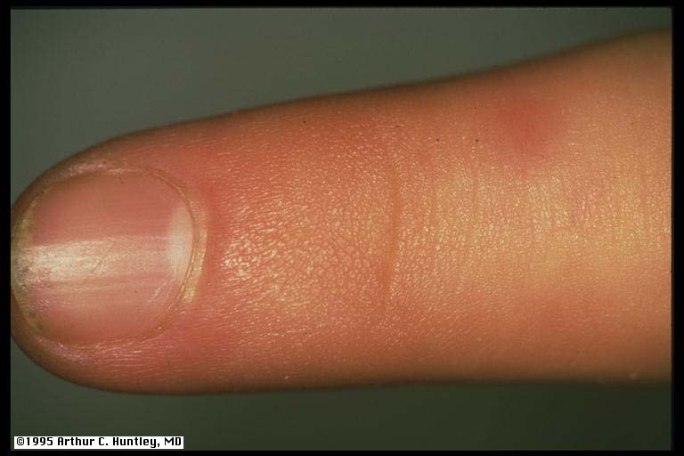
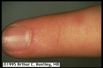
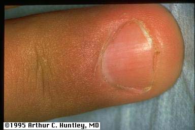
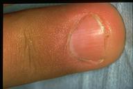
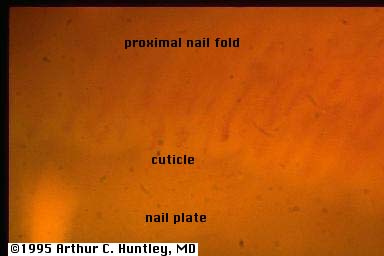
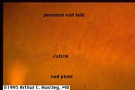
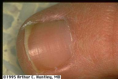
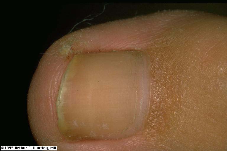
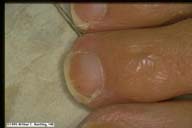
fig3: acanthosis nigricans, proximal nail fold
L M S
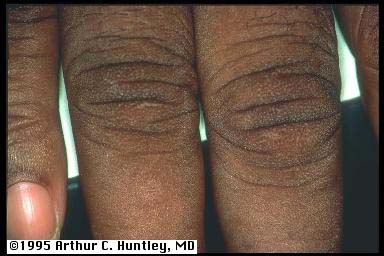
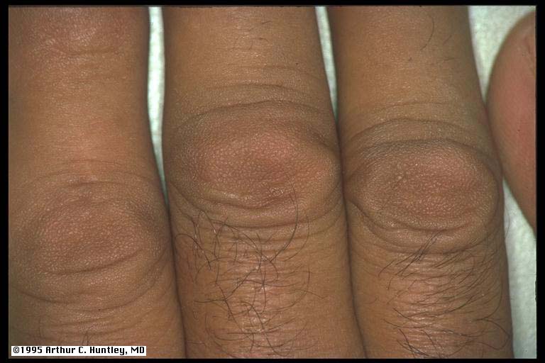
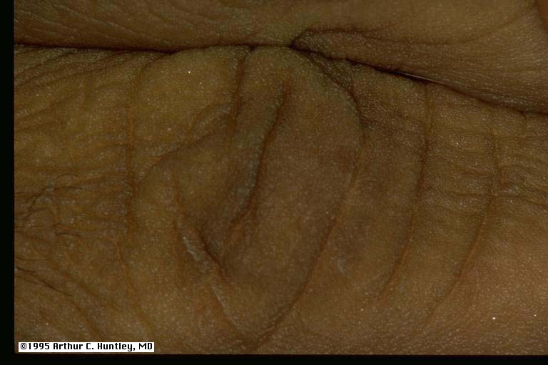
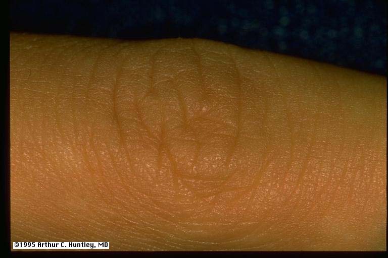
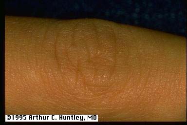
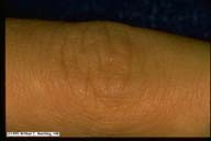
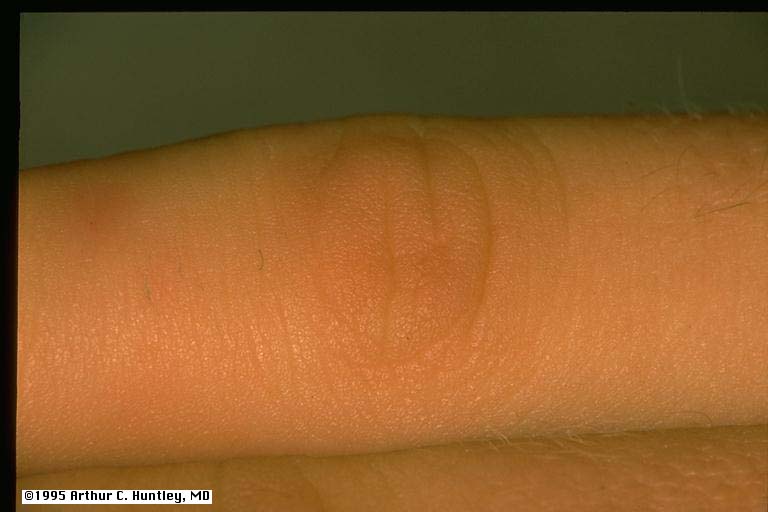
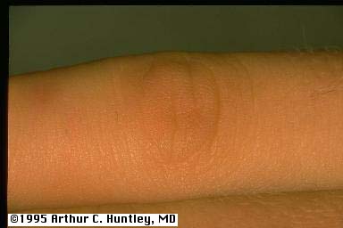
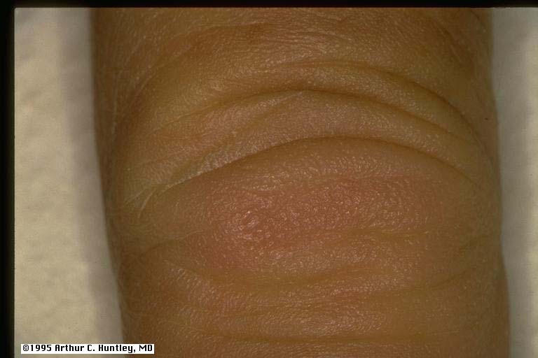
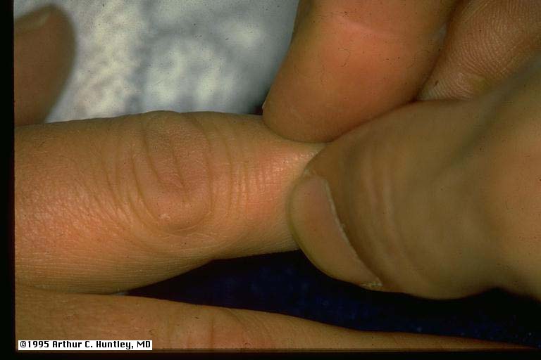
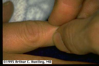
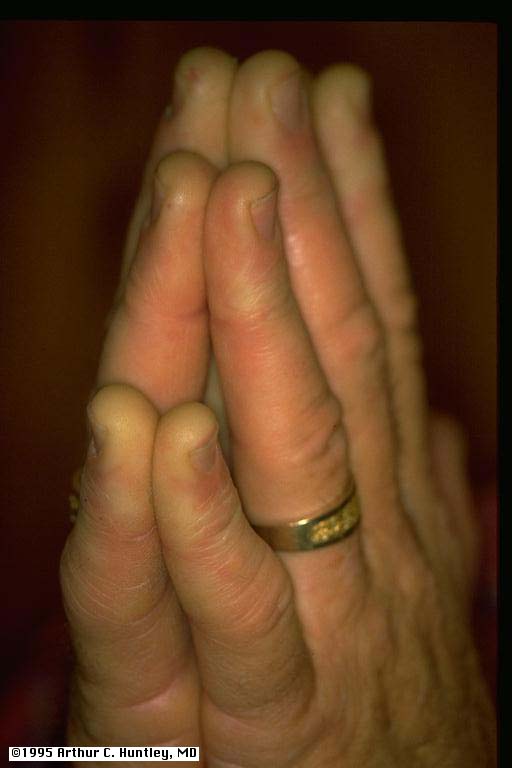
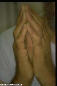
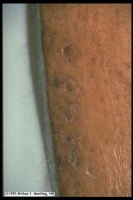
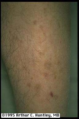
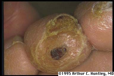
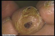
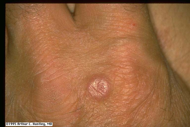
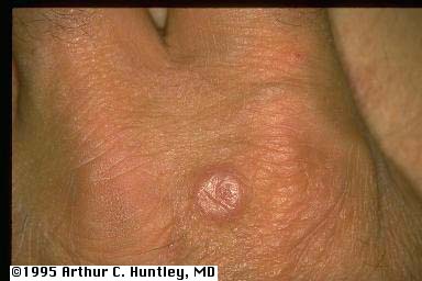
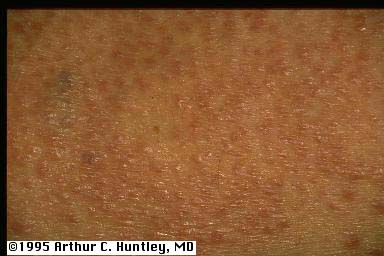
finger pebbles fig4: finger pebbles, knuckle
L M S
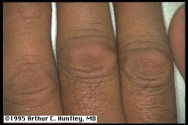
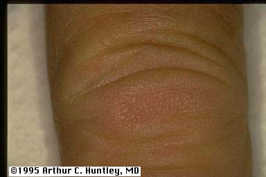
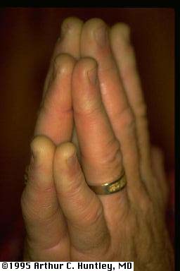
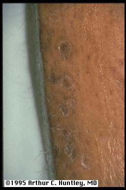
fig 4a: finger pebbles, knuckles
L M S
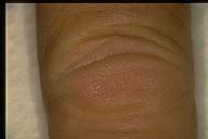
fig 4b: finger pebbles, knuckle
L M S
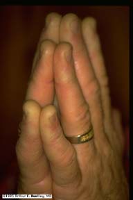
fig 4c: finger pebbles, knuckle
L M S
fig 4d: finger pebbles, knuckle
L M S
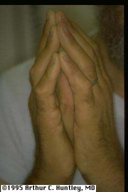
fig 4e: finger pebbles, knuckle
L M S
fig 4f: finger pebbles, knuckle
L M S
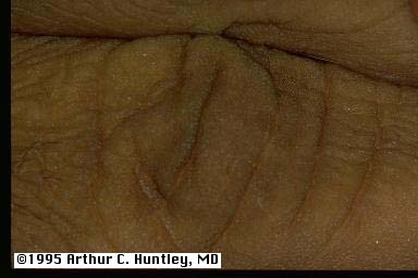
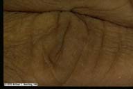
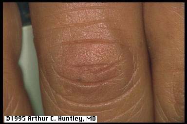
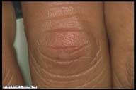
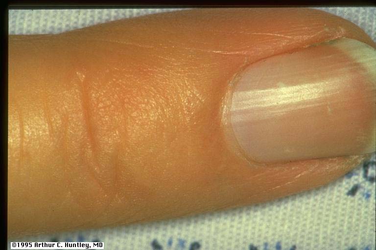
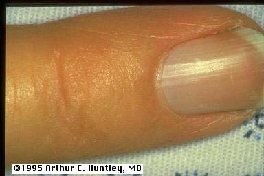
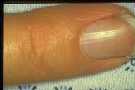
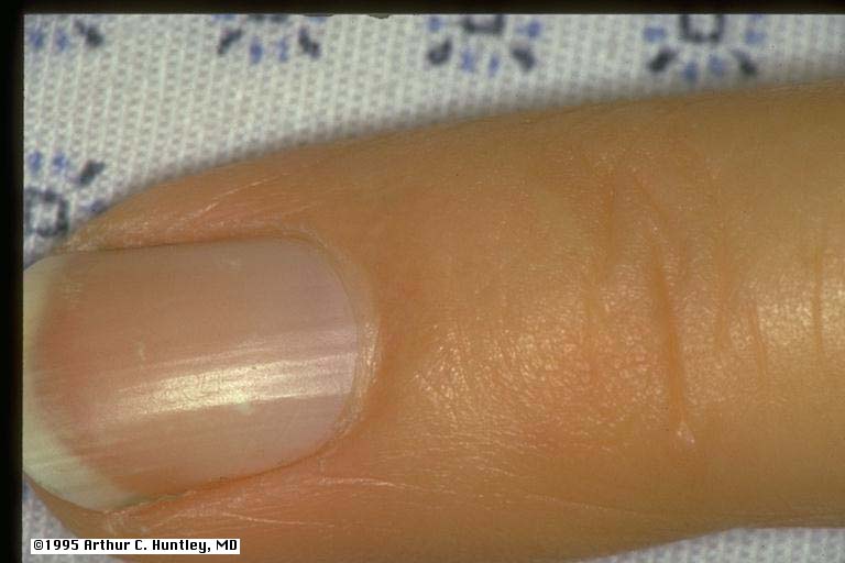
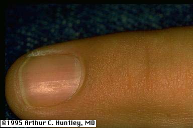
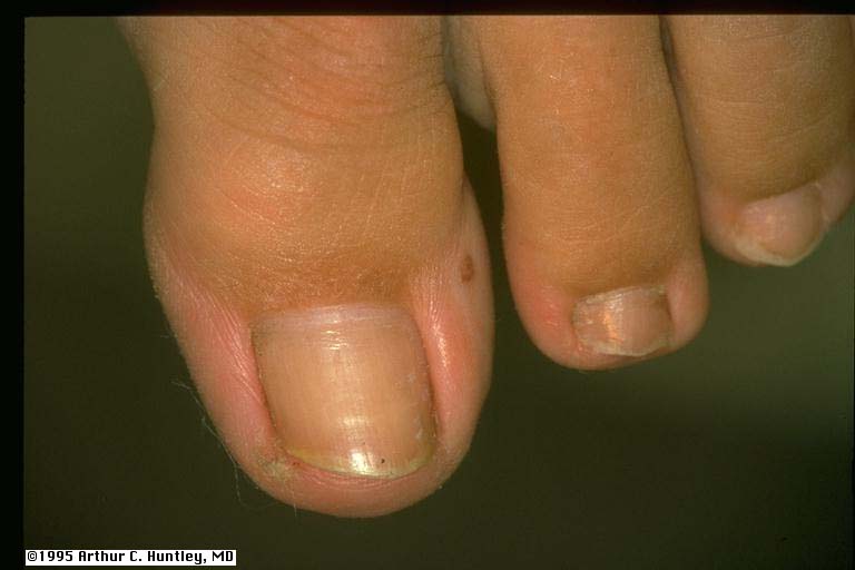
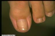
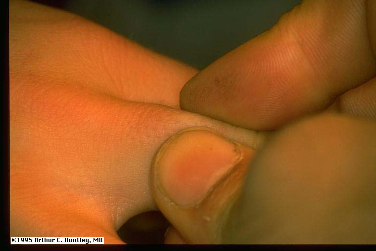
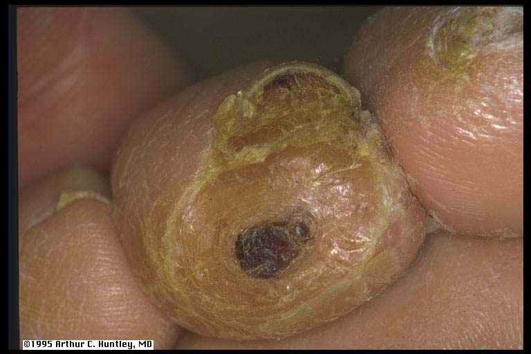
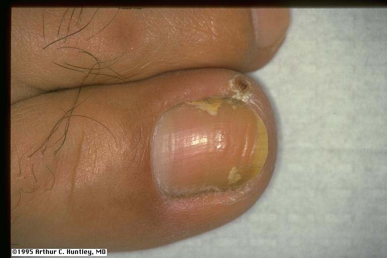
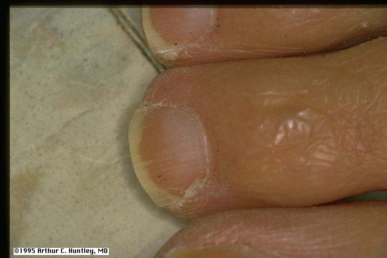
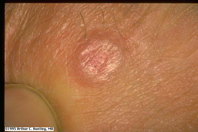
proximal nail fold thickening, pebbles
fig5: finger pebbles, proximal nail fold
L M S
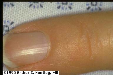
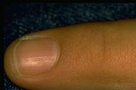
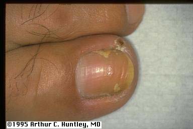
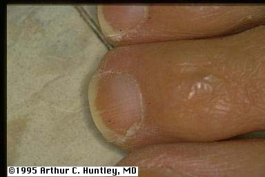
fig 5a: finger pebbles, proximal nail fold
L M S
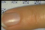
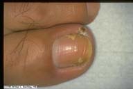
fig 5b: finger pebbles, proximal nail fold
L M S
fig 5c: finger pebbles, proximal nail fold
L M S
fig 5d: finger pebbles, proximal nail fold
L M S
fig 5e: finger pebbles, proximal nail fold
L M S
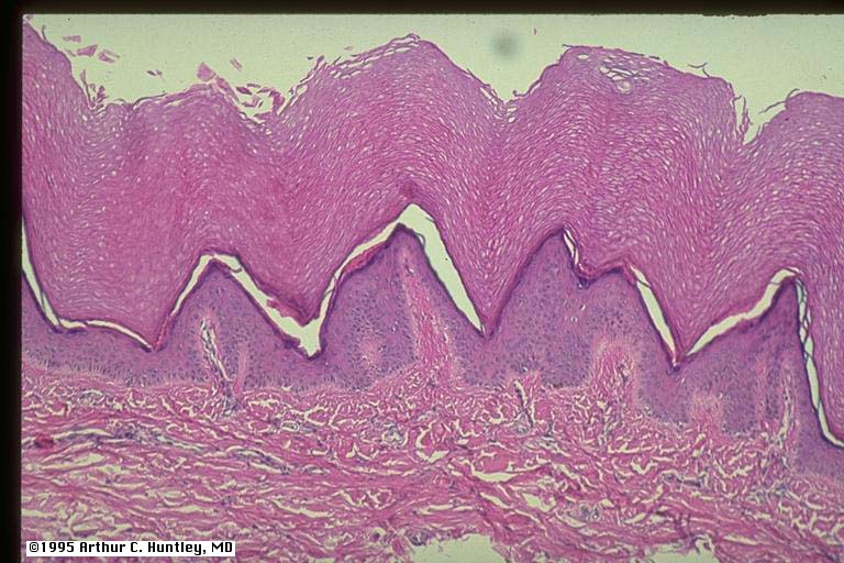
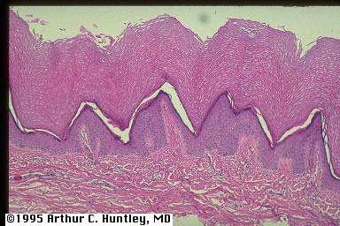
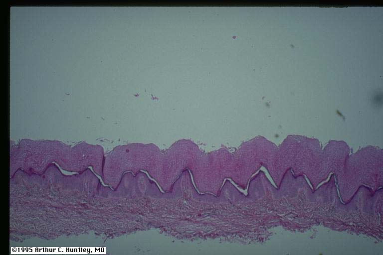
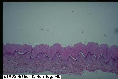
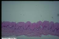
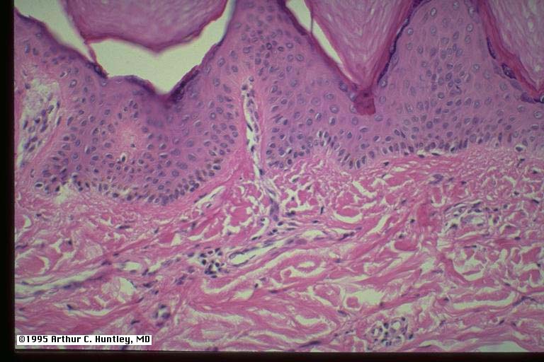
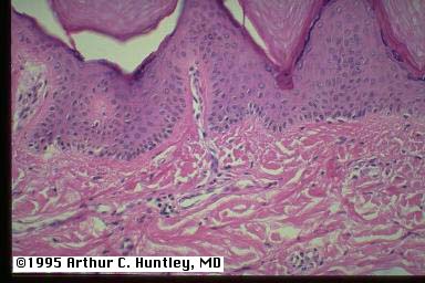
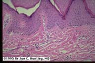
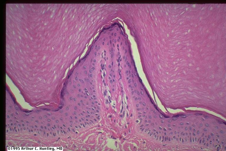
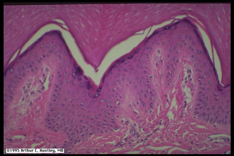
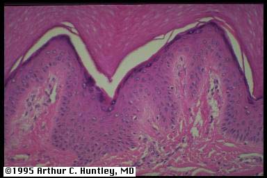
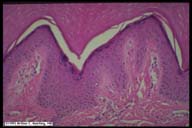
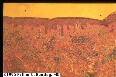
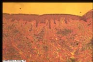
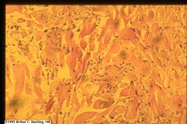
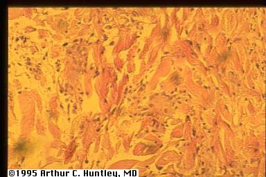
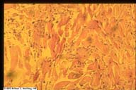
finger pebbles: histology
fig 6: finger pebbles, knuckle skin
L M S
fig 6a: finger pebbles, knuckle skin, low power
L M S
fig 6b: finger pebbles, knuckle skin, medium power
L M S
fig 6c: finger pebbles, knuckle skin, high power
L M S
fig 6d: finger pebbles, knuckle skin, high power
L M S
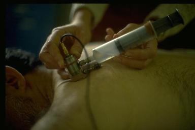
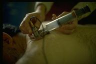
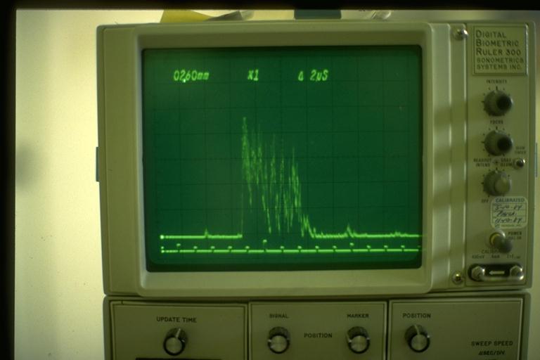
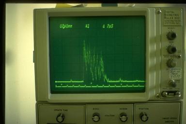
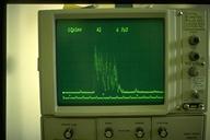
using ultrasound to measure skin thickness
fig7: placement of ultrasound transducer
L M S
fig8: ultrasound display
L M S
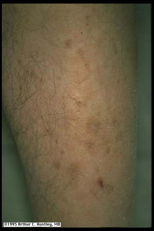
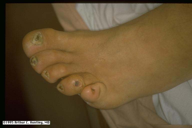
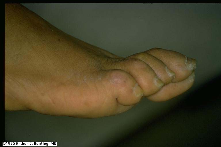
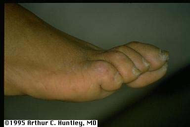
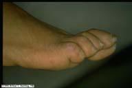
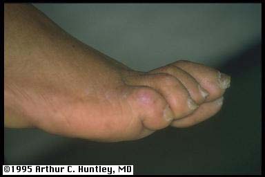
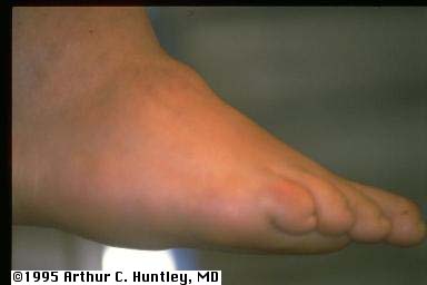
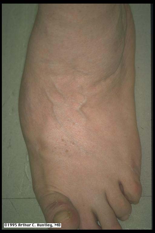
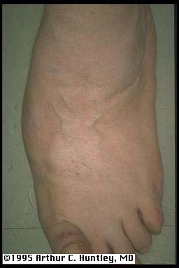
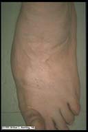
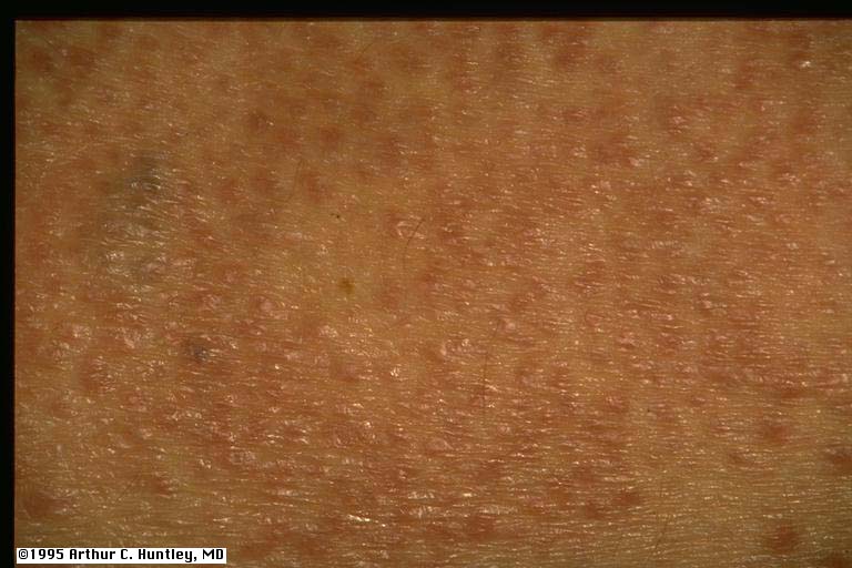
finger pebbles, thick skin on dorsum of foot.
fig9: finger pebbles, knuckle
L M S
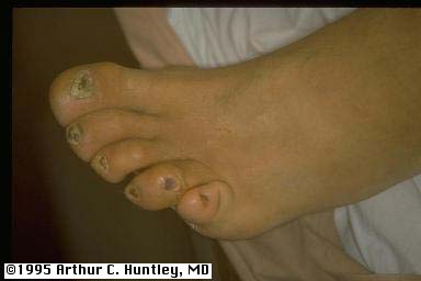
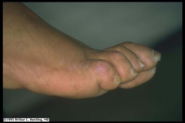
fig 10: pebbles, proximal nail fole of great toe
L M S
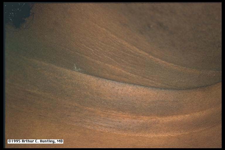
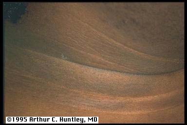
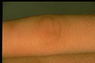
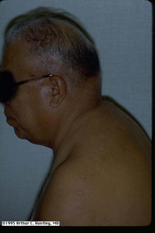
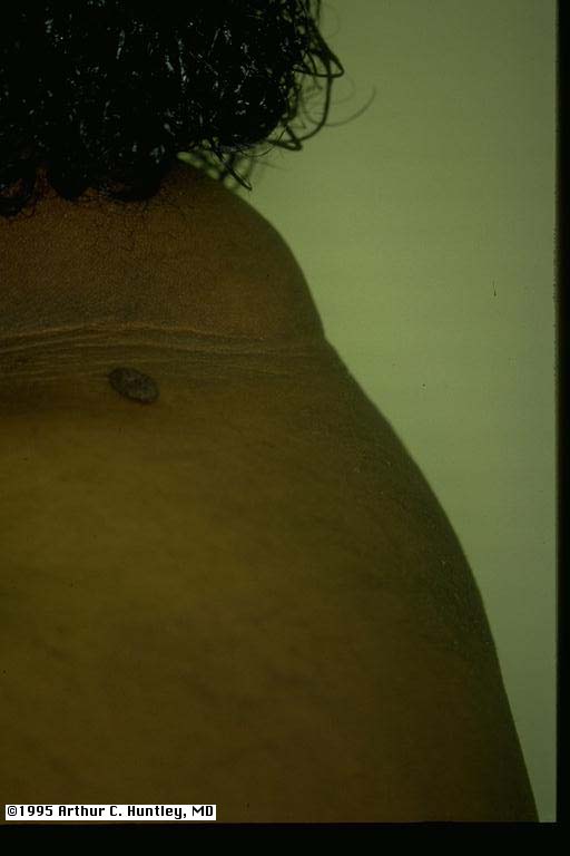
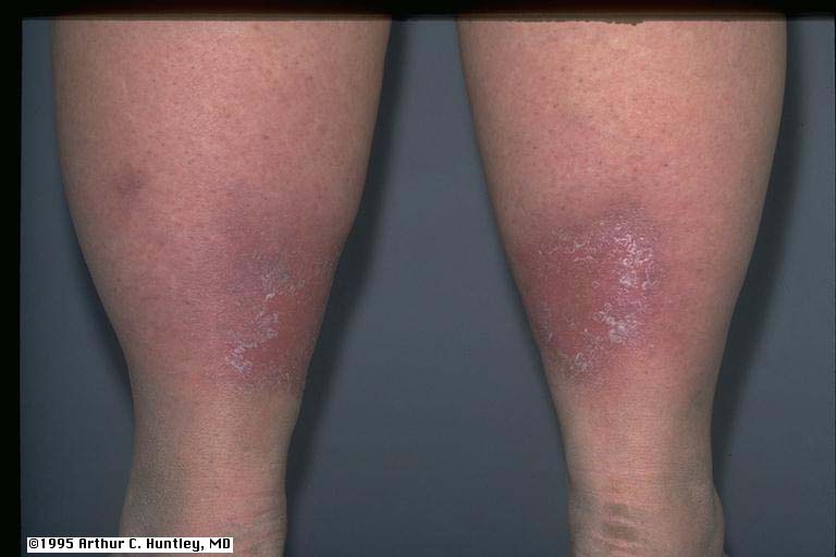
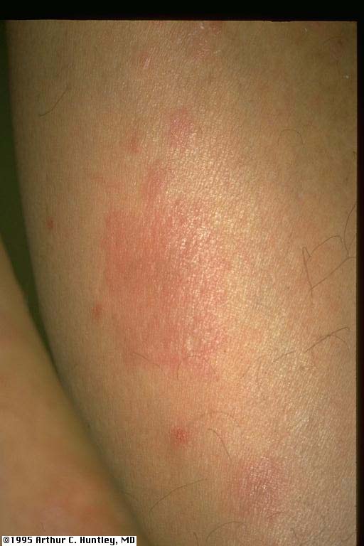
scleredema of diabetes mellitus fig 11: scleredema, back
L M S
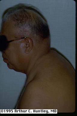
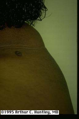
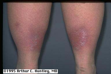
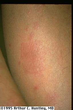
fig 13: scleredema, back
L M S
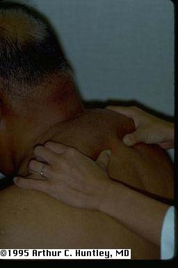
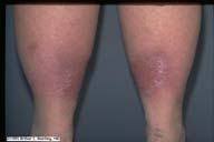
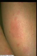
fig 14: scleredema, back
L M S
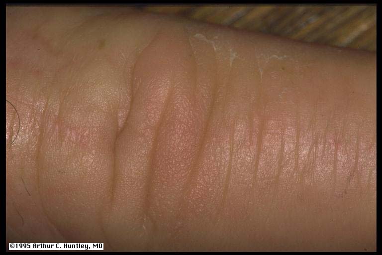
diabetic hand syndrome: thick skin fig 15: taught skin, finger
L M S
fig 16: taught skin, finger
L M S
fig17: taught skin, finger
L M S
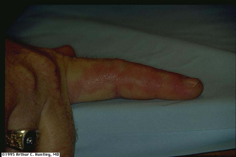
diabetic hand syndrome: joint limitation
fig 18: joint limitation, fifth finger
L M S
fig 19: joint limitation, hands
L M S
fig 20: joint limitation, hands
L M S
fig 22: diabetic nailfold capillaries
L M S
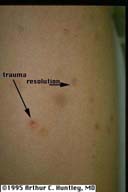
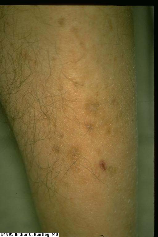
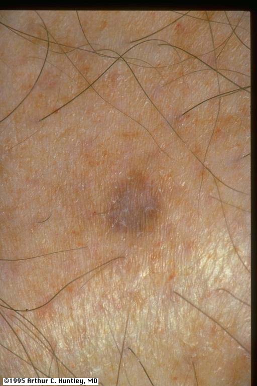
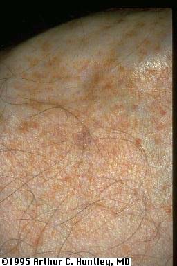
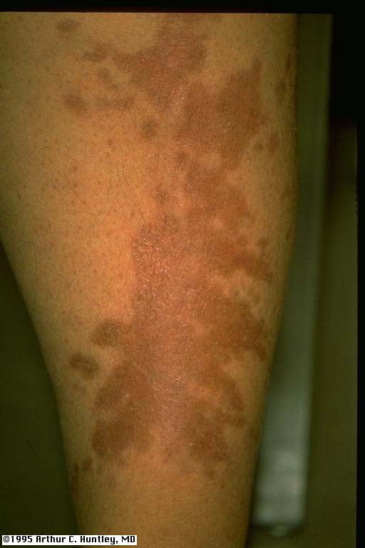
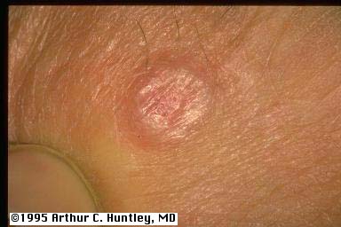
fig 23: diabetic dermopathy
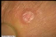
L M S
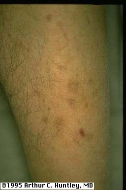
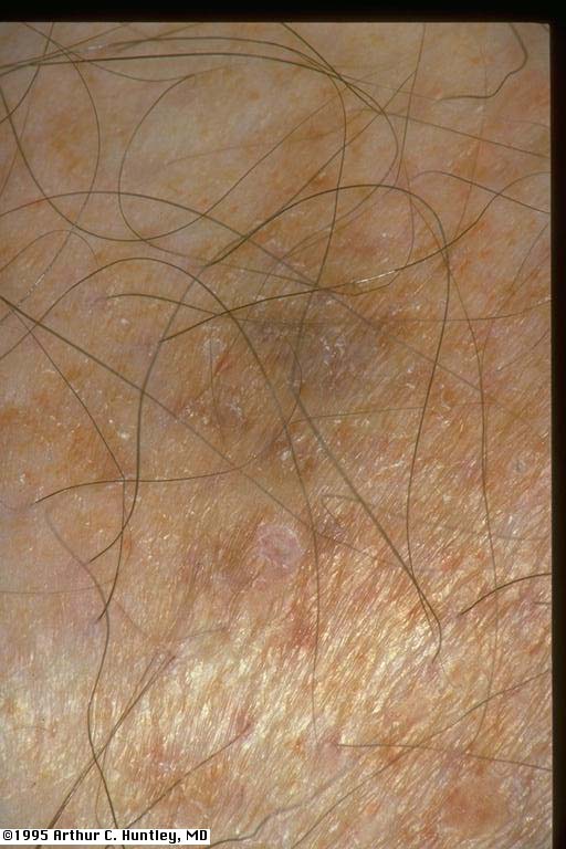
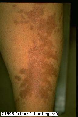
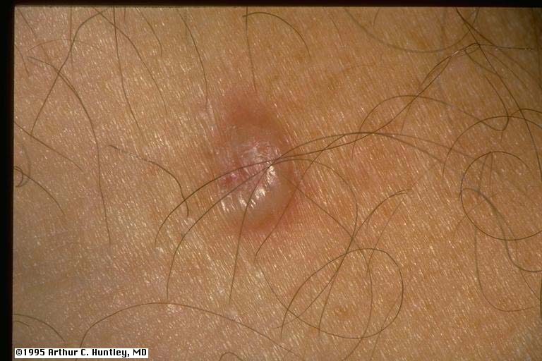
fig 24: diabetic dermopathy
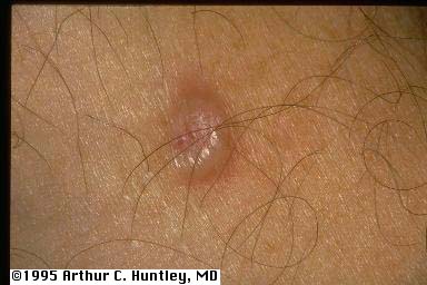
L M S
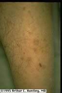
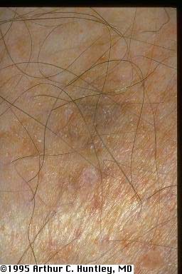
fig 25: diabetic dermopathy
L M S
fig 26: diabetic dermopathy
L M S
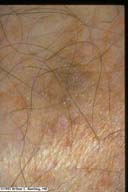
fig 27: diabetic dermopathy
L M S
fig 27a: diabetic dermopathy
L M S
fig 27b: diabetic dermopathy, close
L M S
fig 27c: diabetic dermopathy, close
L M S
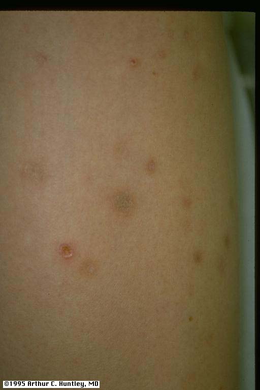
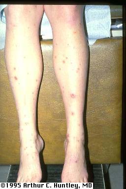
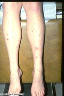
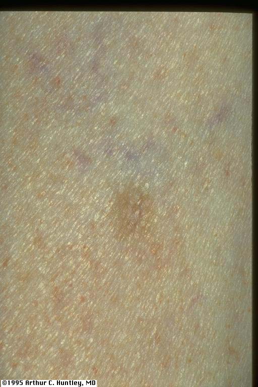
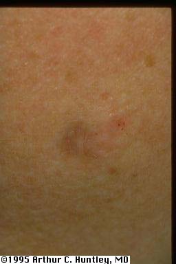
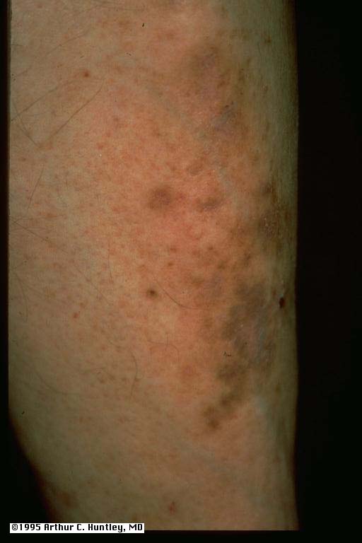
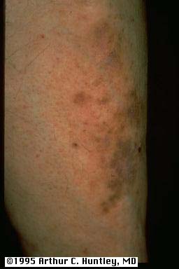
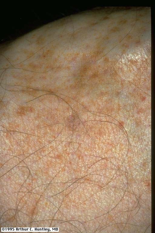
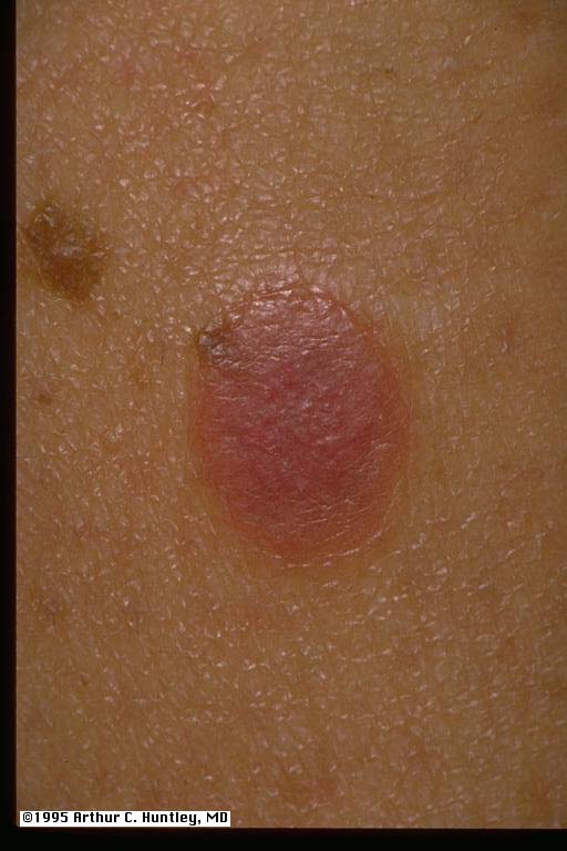
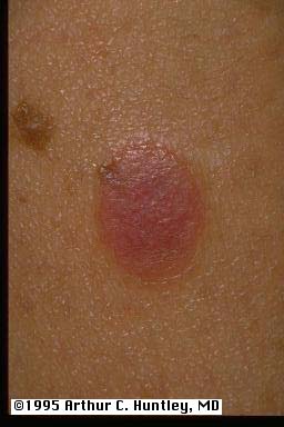
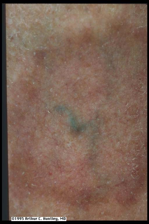
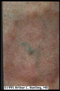
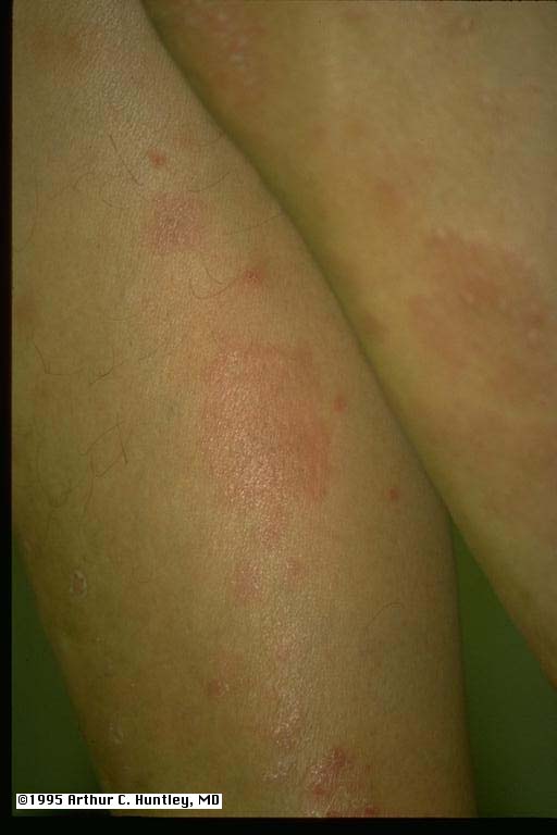
a.pigmented purpura fig 28: pigmented purpura, shin
L M S
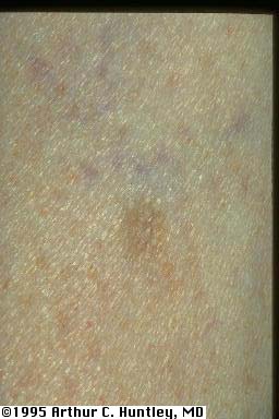
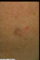
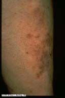
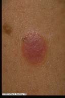
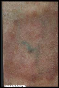
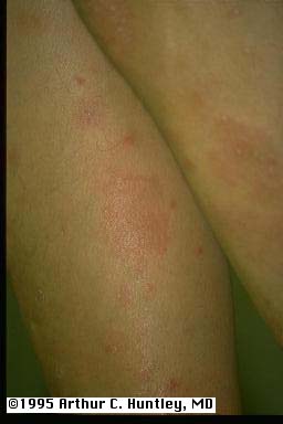
fig 29: pigmented purpura, shin
L M S
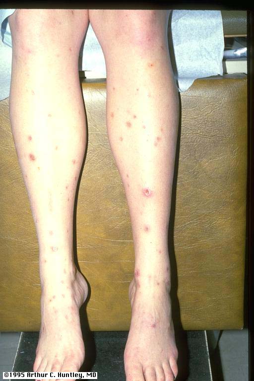
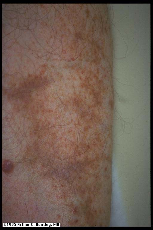
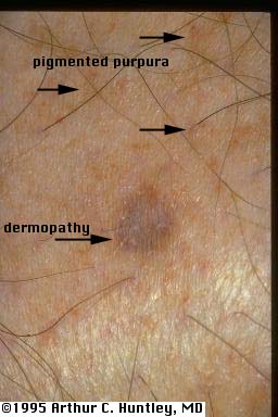
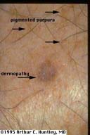
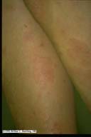
fig 30: pigmented purpura and dermopathy
S M L
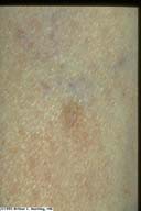
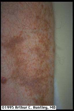
fig 30a: pigmented purpura, close
L M S
fig 30b: pigmented purpura, close
L M S
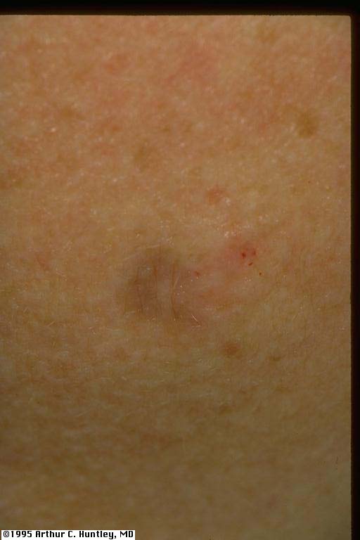
fig 32: sensory neuropathy, erosions on dorsum of toes
L M S
fig 32a: sensory neuropathy, toe ulcer
L M S
motor neuropathy fig33: motor neuropathy, cocked toes
L M S
fig 33a: motor neuropathy, cocked toes
L M S
Charcot foot fig 34: Charcot foot
L M S
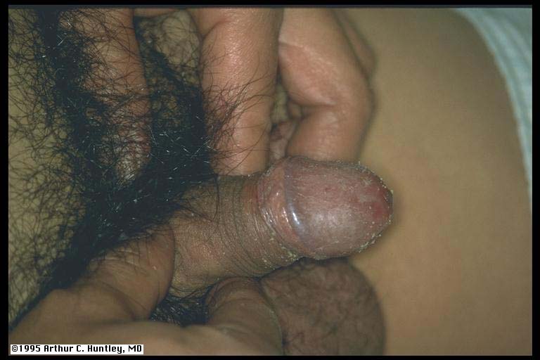
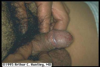
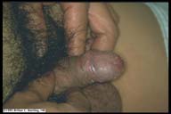
fig 36: Candida infection of glans penis
L M S
fig 37: Candida infection of interdigital space of fingers.
L M S
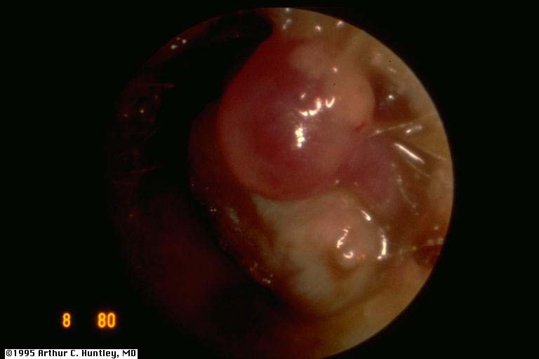
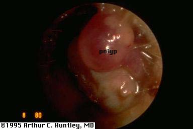
malignant external otitis fig 38: Pseudomonas external otitis with polyp formation
L M S
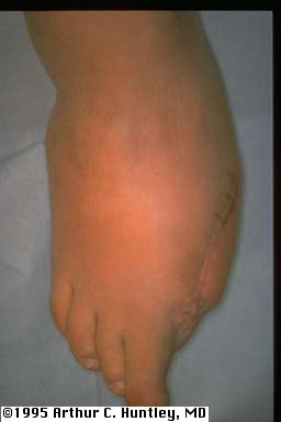
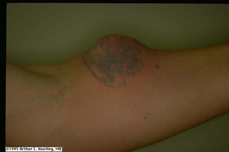
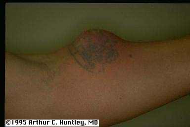
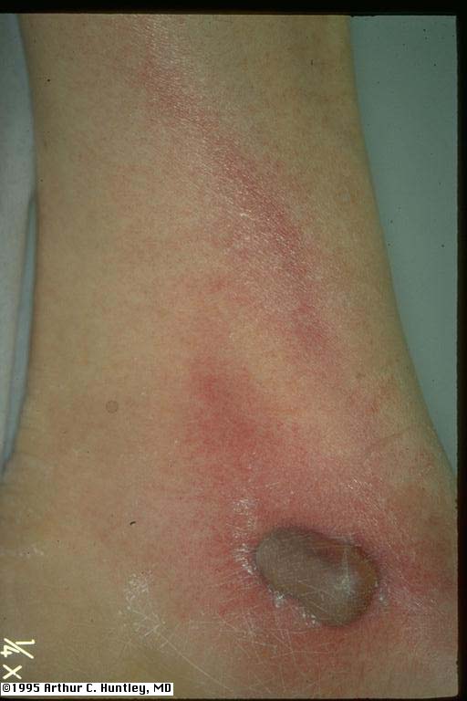
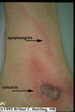
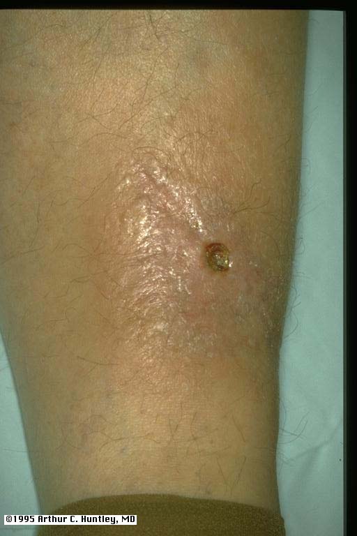
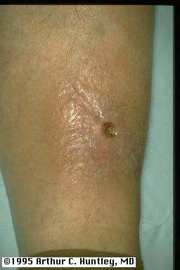
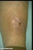
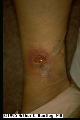
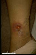
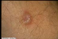
staph and strep fig 39: abscess of forearm
L M S
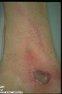
fig 40: leg ulcer with lymphangitis
L M S
fig 41: paronychia and cellulitis/lymphangitis of finger
L M S
dermatophyte infection fig 42: tinea on dorsum of hand
L M S
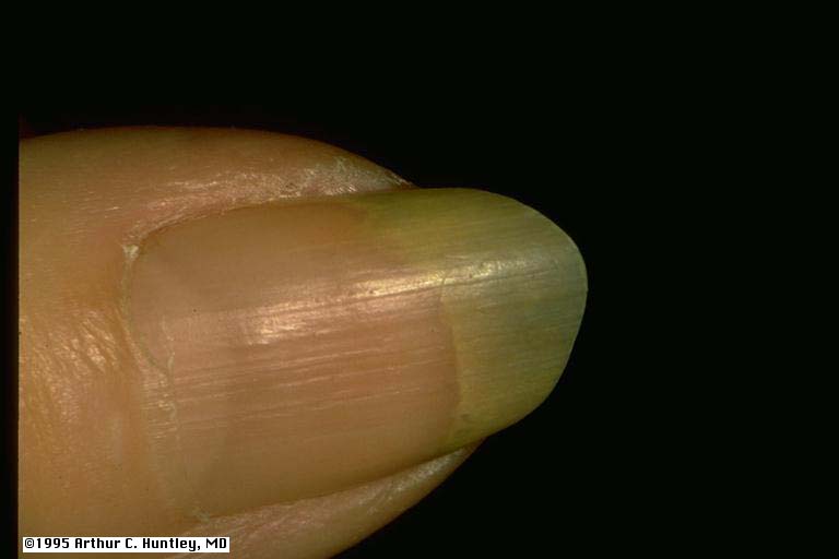
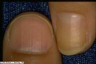
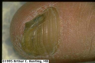
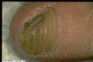
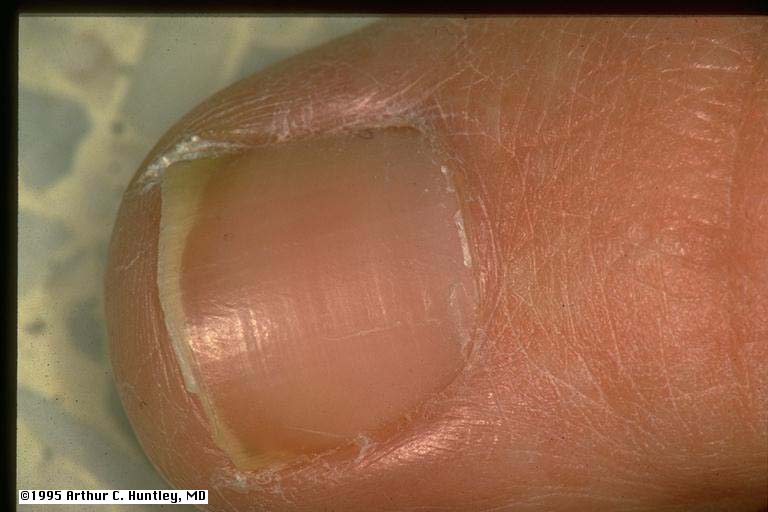
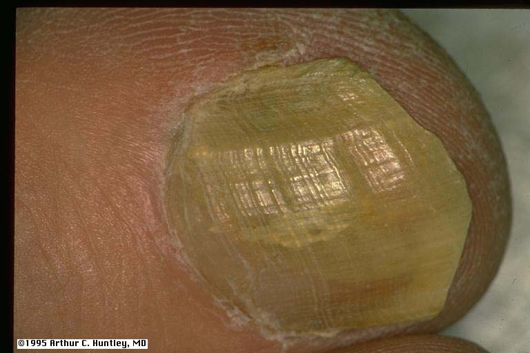
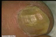
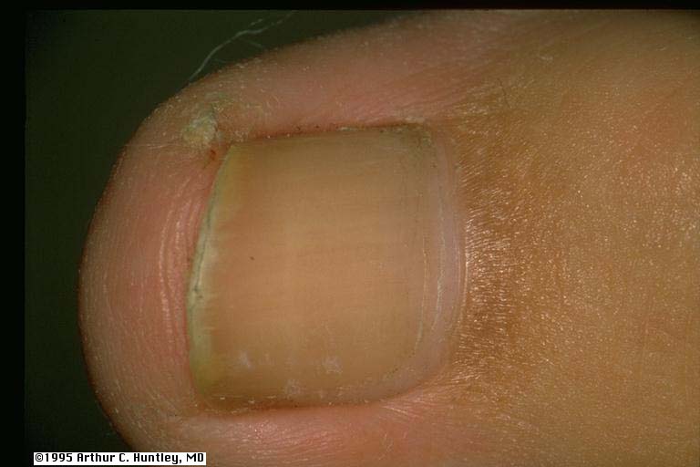
Fig 44: yellow fingernail
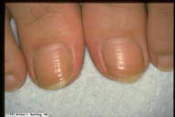
L M S
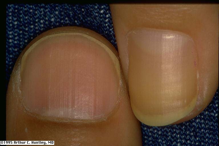
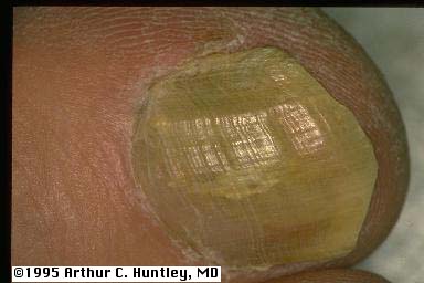
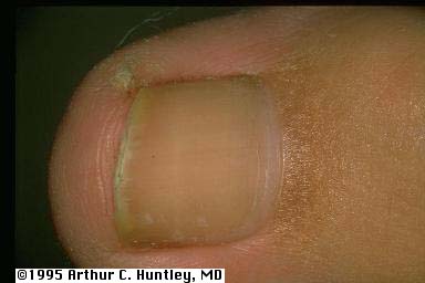
fig 45: yellow vs. normal fingernail
L M S
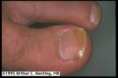
fig 46a: yellow and green discoloration, diabetes, tinea, and pseudomonas
L M S
fig 46b: yellowing of distal aspect of toenails
L M S
fig 46c: yellow/brown discoloration of distal toenail
L M S
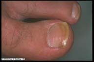
fig 46d: yellow toenail, diabetes and fungus
L M S
fig 46e: yellow toenail, diabetes and fungus
L M S
fig 46f: yellow nail and pebbling of proximal nail fold of hallux
L M S
fig 46g: distal yellowing of toenail
L M S
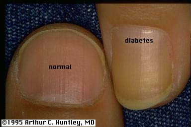
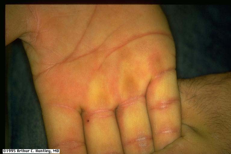
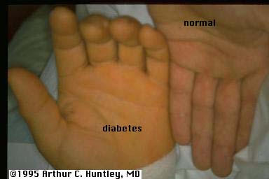
yellow skin fig 47: yellow palm
L M S
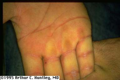
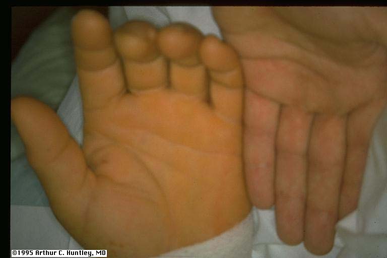
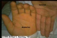
fig 48: yellow vs. normal palm
L M S
fig 49: yellow skin and yellowing of distal nail, hallux
L M S
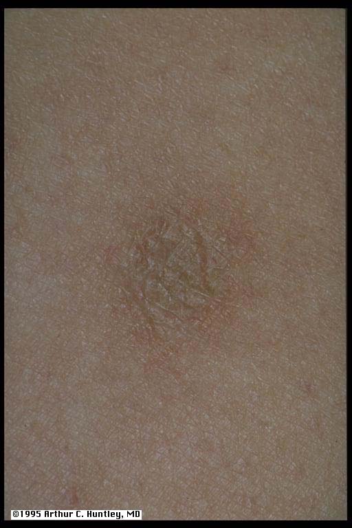
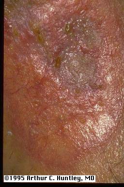
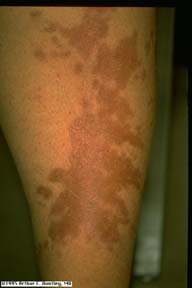
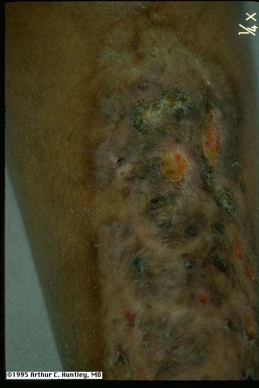
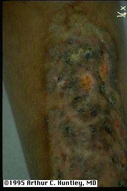
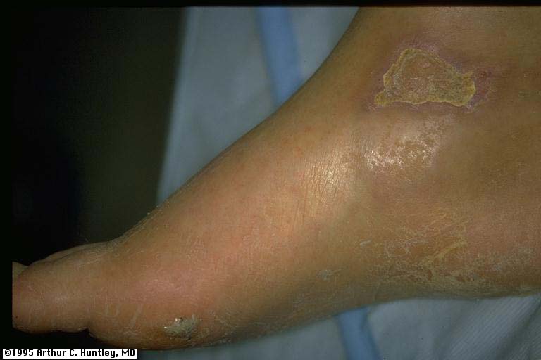
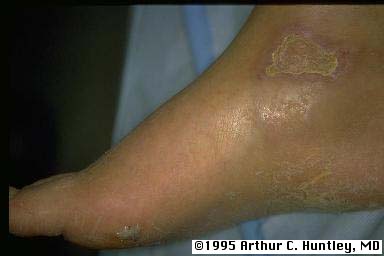
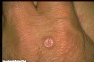
fig 51: necrobiosis lipoidica of legs
L M S
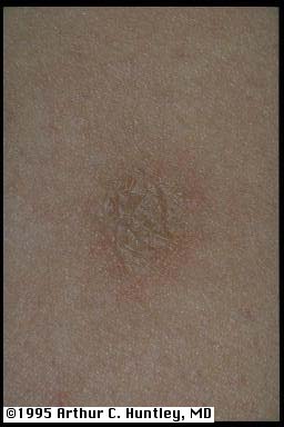
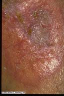
fig 52: necrobiosis lipoidica, early papular lesion, leg
L M S
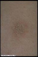
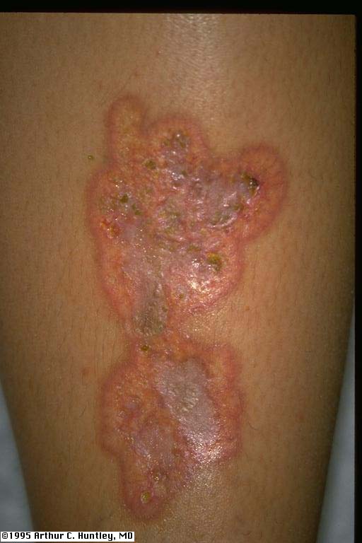
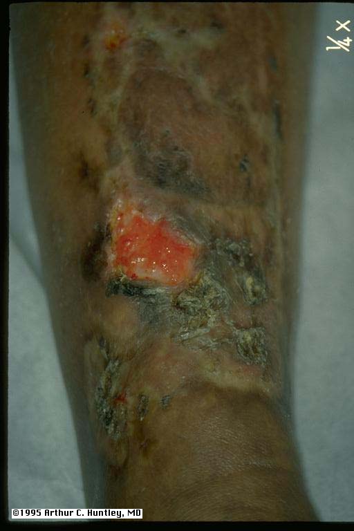
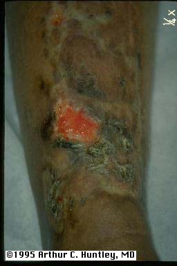
fig 53: necrobiosis lipoidica, leg, close view
L M S
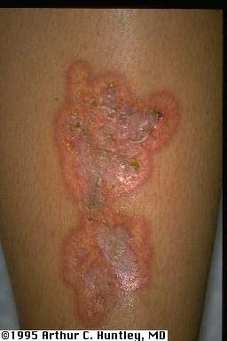
fig 53a: necrobiosis lipoidica, leg, close view
L M S
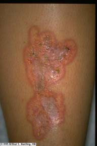
fig 53b: necrobiosis lipoidica, leg, close view
L M S
fig 53c: necrobiosis lipoidica, leg
L M S
fig 53d: necrobiosis lipoidica, leg
L M S
fig 53e:necrobiosis lipoidica, leg, non-diabetic
L M S
fig 53f53e:necrobiosis lipoidica, leg, non-diabetic
L M S
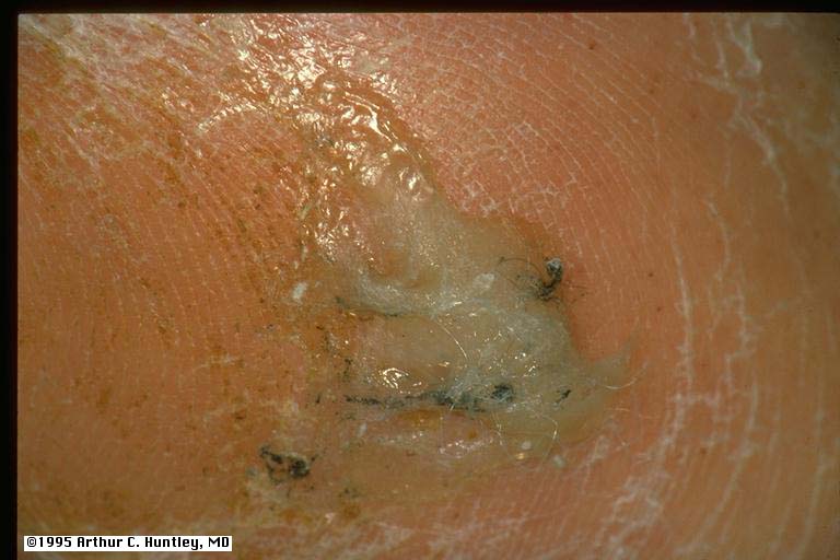
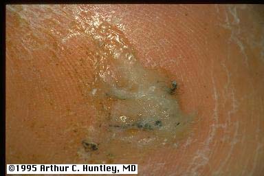
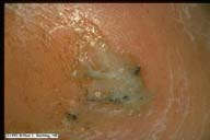
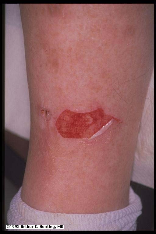
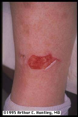
fig 55: diabetic bulla, leg, ruptured
L M S
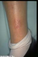
fig 56: diabetic bulla, leg, ruptured with cellulitis
L M S
fig57: diabetic bulla, foot
L M S
fig 58: diabetic bulla, foot, close view
L M S
fig 59: diabetic bulla of leg
L M S
fig 60: erosion of skin by removal of adhesive tape, adjacent to bulla
L M S
fig 62: granuloma annulare, dorsum of hand
L M S
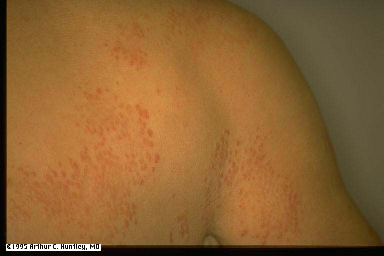
fig 63: disseminated granuloma annulare, back and shoulder
L M S
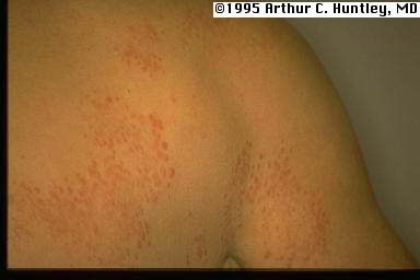
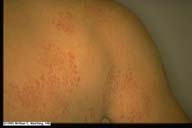
fig 64: disseminated granuloma annulare, back, close view
L M S
fig 64a: granuloma annulare, single lesion
L M S
fig 64b: granuloma annulare, single lesion
L M S
fig64c: disseminated granuloma annulare
L M S
fig 64d: disseminated granuloma annulare
L M S
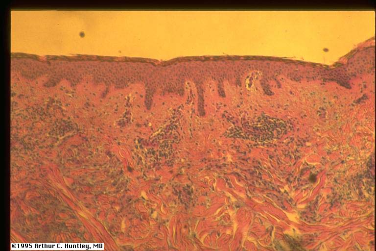
histology of granuloma annulare
fig 65: granuloma annulare, histology
L M S
fig 66: granuloma annulare, histology, close view of dermis
L M S
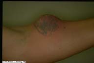
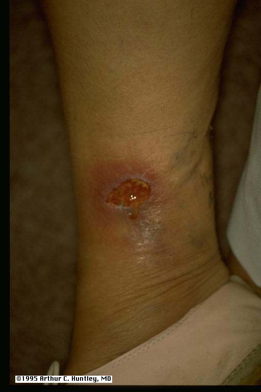
fig 69 (and fig 39): abscess of forearm at site of insulin injection
L M S
All contents copyright © 1995.
Dermatology Online Journal
University of California Davis