Photoessay: The Skin and Diabetes Mellitus
by A Huntley
Dermatology Online Journal, December 1995
Volume 1, Number 2
Granuloma Annulare and Diabetes
Similar to the association of necrobiosis lipoidica and diabetes, it appears that a high percentage of persons with disseminated
granuloma annulare have diabetes mellitus. The individual lesions typically consist of an annular array of erythematous to
brown and slightly translucent papules.
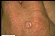
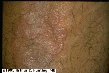
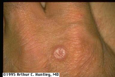
Figs 61,62. Dorsum of the hand of two patients with diabetes mellitus and granuloma annulare. The left-hand image demonstrates
the annular and serpiginous nature of the border. The right-hand image is of an enlarging papule which is developing a central
dell.
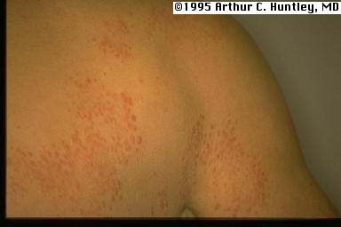
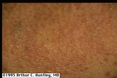
That form which is more associated with diabetes is associated with multiple and widespread lesions.
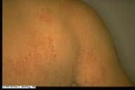
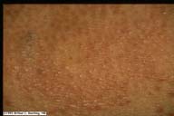
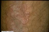
Figs 63,64. Patient with diabetes mellitus who also has disseminated granuloma annulare. The left-hand image demonstrates
the dispersed involvement on the upper back and arm. The right-hand image demonstrates the multiple brown to erythematous
papules on close-up view.
histology
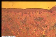
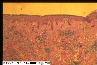
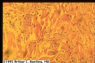
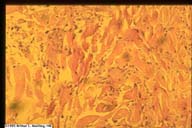 Figs 65,66. Skin biopsy from a patient with diabetes mellitus and disseminated granuloma annulare. The left-hand image demonstrates
both a perivascular and interstitial inflammatory cell infiltrate. The right-hand image demonstrates histiocytes between collagen
bundles.
Figs 65,66. Skin biopsy from a patient with diabetes mellitus and disseminated granuloma annulare. The left-hand image demonstrates
both a perivascular and interstitial inflammatory cell infiltrate. The right-hand image demonstrates histiocytes between collagen
bundles.
additional images of granuloma annulare
All contents copyright (C), 1995.
Dermatology Online Journal
University of California Davis