|
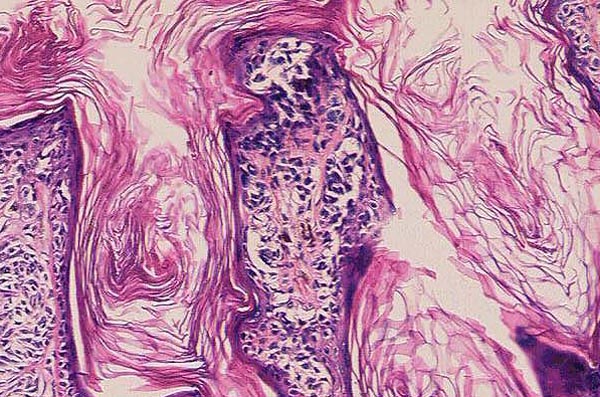
| Figure 5. Atypical melanocytes are seen in both nested and confluent single-cell array at the dermal-epidermal junction. Pagetoid spread is seen throughout the lesion. Atypical melanocytes involve the dermis as well (100X). [Full size view 1641X1219] |
| Return |