Portable, Inexpensive Instruments to Quantify Stratum Corneum Hydration and Skin Erythema: Applications to Clothing Science
Published Web Location
https://doi.org/10.5070/D357r2h4mzMain Content
Portable, Inexpensive Instruments to Quantify Stratum Corneum Hydration and Skin Erythema: Applications to Clothing Science
K J Atkins and M W Thompson
Dermatology Online Journal 7(2): 2
School of Exercise and Sport Science, Faculty of Health Sciences, The University of Sydney, Australia.Abstract
The level of stratum corneum hydration is assessed by measurement of the changes in skin resistance and is referred to as the galvanic skin response or electrical skin resistance. Skin erythema may be assessed by measurement of skin blood flow, tristimulus colorimetry or narrow-band reflectance spectroscoy. Currently available measuring devices are relatively expensive. Presented here are two inexpensive, hand-held, portable instruments which conveniently measure stratum corneum hydration and skin erythema.
Introduction
The degree of skin blood flow and stratum corneum (SC) hydration in response to clothing are associated with the perception of fabric comfort. [1] The SC hydration and skin redness (erythema) have been measured previously to assess how these skin properties influence perceived comfort to different types of clothing [1]. The level of stratum corneum hydration is mainly inferred from changes in skin resistance, referred to as the galvanic skin response, electrical skin resisitance or electrodermatographic result. [2,3] Previous investigations assessing clothing comfort have measured skin blood flow using laser doppler methods. [1] Other methods available to quantify skin redness (erythema), used mainly in the clinical setting, include tristimulus colorimetry and narrow-band reflectance spectroscopy. [4,5,6]
While measurement systems are available to quantify SC hydration [5,7,8] and skin erythema [4,6,9] there remains a paucity of information on the effect of different thermal environments and fabric properties on these skin parameters [1]. Current measurement systems are relatively expensive which may account for the choice of investigators not to include the measurement of these skin properties in their studies. We present here two handheld instruments that we have used to measure stratum corneum hydration (inferred from skin resistance) and skin erythema that provide an inexpensive, portable alternative to current instruments that can be constructed by researchers. These devices were approved by the The University of Sydney Electronics Engineering-Research Support department and Human Ethics Committee and were found to comply with Australian safety standards [10].
Skin Resistance Monitor: An Instrument to Measure SC Hydration
 |
| Figure 1 |
|---|
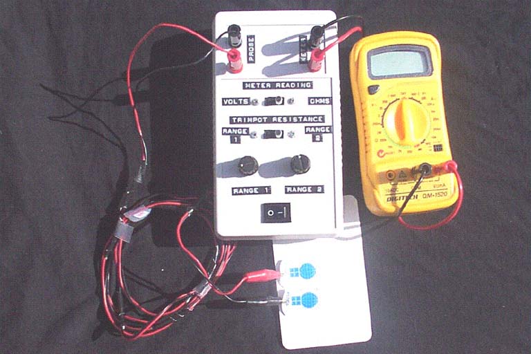
The skin resistance monitor is a device powered by a nine volt battery designed to measure skin resistance using a wheatstone bridge circuit. The monitor is shown in Figure 1. The measure of skin resistance is used as an indicator of changes in the moisture content of the skin.
Materials and Methods
Probe leads and Electrode Attachment
A small adhesive backed electrode with conductive gel (Kendall Care™610 resting ECG electrode) was used successfully with this device that adhered well under high sweating rate conditions. The advantage of using this electrode is that it can be applied to the skin surface without having to prepare the skin, which could alter skin erythema readings, prior to its attachment. The probe leads were constructed of insulated hook up wire, 2.85 m in length. The probe leads were attached to the electrodes using mini-insulated alligator clips. Surgical tape (Transpore™) was used to secure the electrodes and attached alligator clips to the skin surface. The probe leads were attached to the wheatstone bridge circuit via a standard banana plug and low profile banana socket arrangement.
The Wheatstone Circuit
 |
| Figure 2 |
|---|
The circuit diagram is shown in Figure 2. The circuit was constructed on Velstra Strip Board and housed in a plastic case of dimensions 180 mm x 100 mm x 40 mm. The hand held case provided a tongue and groove sealing system to minimise the entry of sweat into the device. All electrical components were attached to the strip board using general purpose solder (1.25 mm 60/40 tin/lead).
Resistances R1 and R2 were 1 kΩ with 1% tolerance. R3 was used to balance the wheatstone bridge. Two ranges were available, selected using a DP3T break before make slide switch, to improve adjustment of R3 to balance the circuit. Range 1 consisted of a 500 kΩ potentiometer (24mm rotary pot, linear taper 0.5W 500V). Range 1 was suitable when the skin resistance was relatively high under conditions of minimal moisture within the skin. Under high sweating rates it was difficult to balance the wheatstone circuit with Range 1 as the skin resistance values decreased with greater moisture content in the skin. Range 2 was then included in this device for finer adjustments to balance the circuit. Range 2 was comprised of a 100 kΩ potentiometer with a ten turn adjustment range (Spectrol model 534, LIN ±0.25%, ±5%).
Voltage and resistance readings were measured using a high impedance multimeter (Digitech QM 1520). A DP3T break before make slide switch was used to change between the voltage and resistance meter readings. The meter leads consisted of medium duty insulated hook-up wire which were attached to the monitor using a standard banana plug and low profile banana socket arrangement.
Calibration of the Skin Resistance Monitor
The skin resistance monitor was calibrated through attaching the probe leads to resistors (Metalfilm resistors, 1/4W, 1% tolerance) ranging in value from 1 kΩ to 500 kΩ.
Methods Used to Operate the Skin Resistance Monitor
After balancing the wheatstone bridge, R3 is equal to the skin resistance provided that the internal resistance within the battery and the circuit does not confound the resistance measure. This was achieved by removing power from the circuit and disconnecting the probe lead from the monitor prior to measuring R3. The following steps were used to operate the monitor:
- 1. The probe leads were attached into the circuit.
- 2. The circuit was turned on.
- 3. The slide switch was placed in the 'voltage' position.
- 4. The multimeter was switched to read volts.
- 5. The appropriate range for R3 was selected using the slide switch.
- 6. The circuit was balanced by adjusting the potentiometer of the given range selected.
- 7. After the circuit was balanced, the circuit was turned off.
- 8. The probe lead (either positive or negative) was disconnected from the monitor.
- 9. The slide switch was placed to the 'ohms' position.
- 10. The multimeter was switched to read resistance.
- 11. The resistance value of R3 was determined.
Skin Erythema Monitor
 |
| Figure 3 |
|---|
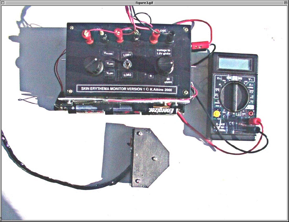
The skin erythema monitor quantifies changes in skin redness based on the amount of light reflected (measured in luxes) from the skin surface from a known amount of illuminated light delivered to the skin surface. A reduction in the luminous flux from the skin surface was used as an indicator of a darker skin surface as a result of an increased skin blood flow within the measurement region. The monitor is shown in Figure 3.
Materials and Methods
 |
| Figure 4 |
|---|
Construction of the Skin Erythema Monitor
The globe and LDR electrical circuits were constructed separately on Velstra Strip Board and were placed in a plastic project box of dimensions 13 mm x 7 mm x 33 mm. The connection of the multimeter and device probe to the circuits was achieved through standard banana plug and low profile banana socket connections. All electrical components were connected using general use solder (1.25 mm 60/40 tin/lead).
The device probe was constructed of balsa wood, 3 mm thick. Insulated spacers 2 cm in length were used to provide structural support to the probe. The probe leads were made of insulated hook-up wire.
Control of Illumination of the Skin Surface
 |
| Figure 5 |
|---|
The device probe is shown in Figure 4. The electrical circuit for the probe is shown in Figure 5. An isotropic light source (where light is radiated equally well in all directions) was used consisting of a spherical panel mount globe (6.3V 300 mA). The globe was situated 2.5 cm from the skin surface. The use of a spherical globe ensured that the luminous intensity delivered to the skin surface was constant. To prevent additional light reflected from the inside and outside surfaces of the probe to the skin surface the probe surfaces were painted in matt black (Humbrol) except for the probe chamber that housed the globe which was painted gloss silver (Humbrol) to increase the intensity of light delivered to the skin surface. Light was delivered to the skin surface through a 1 cm2 aperture at an angle of 45°. The probe was placed directly on the skin to prevent ambient light from influencing the measurement.
The globe circuit was powered by a 4.5 V battery source consisting of three 1.5 V batteries. To reduce the variability in voltage across the globe the batteries were secured firmly together using AA battery holders mounted to the monitor case. The globe circuit was constructed separate to the light dependent resistor (LDR) circuit to further reduce the potential variation in voltage delivery to the globe that the components within the LDR circuit might have had. The voltage across the globe was set at 3.20 V, adjusted using a 30 Ω variable resistor. Voltage measures were obtained using a multimeter (Dick Smith Electronics Q-1419).
The luminous flux emitted from the globe was measured using a LDR (Dick Smith Electronics DSCD01). The relationship between illumination and resistance of the LDR is given by:
R = A·L-0.85 (equation 1)
where:
R = resistance of the light dependent resistor in Ω
A = a constant (~ 340 x 103)
Electrical Circuit for the LDRs and Calculation of Reflected Light from the Skin Surface
The circuit for the LDRs was powered by a 9V battery. LDR1 was used to measure the illumination of the globe to check for variation in the emitted light from the globe. LDR2 measured the amount of light reflected from the skin surface. A toggle switch was used to change between LDR1 and LDR2. The LDRs were part of a voltage divider circuit to provide an output voltage (Vo) with changes in illumination. The fixed resistor was 250 Ω. The resistance of the selected LDR (RLDR) was calculated from:
RLDR = ((Vi x 250 Ω)/Vo)/ 250 Ω (equation 2)
where:
Vo = output voltage
Vi = input voltage to the LDR
RLDR = the LDR resistance (Ω)
 |
| Figure 6 |
|---|
| Figure 6 shows the standard reference colours used of known surface luminosity to assess the reliability and repeatability
of the skin erythema monitor. The reference surfaces were produced using the colours function in Microsoft® Excel. The relationship
between the output voltage readings (mean ± SD) in response to changing resistance in LDR2 versus each reference surface is
shown in Figure 7. Changes within the output voltage produced by an altered resistance in LDR2 demonstrated a consistent curvilear
response to increasing luminosity of the reference surfaces (Figure 7). The degree in variation of repeated measures for a
given reference surface was low. |
With this circuit configuration, Vodecreased as illumination of the LDR was reduced. Measures of Vo, Vi and RLDR were obtained using a multimeter (Dick Smith Electronics Q-1419). A rotary break before make switch (2 pole 5 positions) was used to change between Vofor the globe circuit, and Vo, Vi and RLDR meter readings from the LDR circuit. After RLDR was determined, the amount of light reflected from the skin surface was calculated using equation 1.
Reliability and Repeatability of the Skin Erythema Monitor and Example Data
 |
| Figure 7 |
|---|
 |
| Figure 8 |
|---|
| Figure 8 is an example of skin surface luminous flux data collected during a human wear trial assessing the effect of different types of fabric on skin erythema. |
Methods Used to Operate the Skin Erythema Monitor
The following steps were used to measure reflected light from the skin surface:
- 1. The multimeter was set to read volts.
- 2. The globe and LDR circuits were switched on just prior to taking measurements.
- 3. The Vi position of the rotary switch was selected to record the voltage across the LDR.
- 4. The Vglobe position of the rotary switch was selected. The voltage across the globe was set at 3.20 V.
- 5. The probe was held firmly against the skin at the measurement site.
- 6. The VLDR position of the rotary switch was selected. The voltage across LDR1 and LDR2 was measured (Vo). LDR1 and LDR2 were selected using the DP3T slide switch.
- 7. After Viand Voacross LDR2 were measured the globe and LDR circuits were switched off.
- 8. The luminous flux from the skin surface was calculated using equation 2.
Discussion
The skin resistance monitor is intended to quantify changes in the moisture content of the skin caused by diffusion of moisture into the stratum corneum from underlying tissues, active sweating and water absorbed from the external environment [11]. However, the presence of capillary blood flow could also influence changes in skin resistance. To interpret measures of skin resistance, we have previously measured both the local sweating rate and skin erythema to determine the contribution of sweat within the skin versus skin blood flow to the overall skin resistance measure. Local sweating rate can be measured through dew point hygrometry methods [12].
The probe for the skin erythema monitor was constructed to provide a constant illumination of the skin surface and the measure of reflected light from the 1 cm2 illuminated skin area. This was attempted by placing the probe directly on the skin and painting the probe surfaces black to reduce the confounding affect of other sources of reflected light. However, the skin is translucent and there is a possibility that ambient light could enter the measurement site via skin areas not covered by the probe [3]. In our studies the skin area around the probe was covered by clothing and access for the probe to the measurement site was gained through an opening in the clothing (covered by a fabric patch) with the same dimensions as the probe to minimise the entry of ambient sources of light.
A further source of variation in luminous flux from the skin surface is the presence of moisture on the skin surface that acts to increase the amount of reflected light. Drying the measurement site prior to taking a measure will reduce this confounding affect.
Acknowledgments
The authors wish to acknowledge Tim Turner and Dr Barry Holcombe from the School of Exercise and Sport Science, The University of Sydney, and John Eisenhuth from the University of Sydney Electronics Engineering-Research Support department for their assistance in the development of our devices. The authors are grateful for the financial assistance from the 'Woolmark' company to fund this work, and to Michael Harries from 'tyco Healthcare' for his advice on an appropriate electrode for use with the skin resistance monitor and the provision of electrodes. We also thank the staff at 'Dick Smith Electronics' for assisting us in the selection of required electrical components.
References
1. Hatch KL, Markee NL, Prato HH, Zeronian SH, Maibach HI, Kuehl RO, Axelson RD, In Vivo cutaneous response to fabric. Part V: Effect of fibre type and fabric moisture content on stratum corneum hydration. Text Res J 1992;62(11):638-47.2. Korr IM, Thomas PE, Wright HM. Patterns of electrical skin resistance in man. J Neural Transmission 1958;17:77.
3. Shriver MD, Parra EJ. Comparison of narrow-band reflectance spectroscopy and tristimulus colorimetry for measurements of skin and hair color in persons of different biological ancestry. Am J Phys Anthropol 2000;112(1):17-27. PubMed
4. Kollias N, Baqer AH. Quantitative assessment of UV-induced pigmentation and erythema. Photodermatol 1988;5(1):53-60. PubMed
5. Potts RO, Stratum corneum hydration: experimental techniques and interpretations of results. J Soc Cosmet Chem 1986;37:9-33.
6. Seitz JC, Whitmore CG, Measurements of erythema and tanning responses in human skin using a tristimulus colorimeter. Dermatologica 1988;177:70-5. 7. Jacques SL, Maibach HI, Susskind C. Water content in stratum corneum measured by a focussed microwave probe: normal vs psoriatic. Bioengin Skin Newslet. 1981;3:118.
8. Triebskorn A, Gloor M, Greiner F. Comparative investigations on the water content of the stratum corneum using different methods of measurement. Dermatologica 1983;167(2):64-9. PubMed
9. Diffey BL, Oliver RJ, Farr PM. A portable instrument for quantifying erythema induced by ultraviolet radiation. Br J Dermatol 1984;111(6):663-72. PubMed
10. AS 2500, Appendix E: Guidelines for the use of electrically operated physical therapy equipment, stimulators and similar energy delivering devices. pp. 47-48, 1986.
11. Hatch KL, Wilson DR, Maibach HI, Fabric-caused changes in human skin: In vivo stratum corneum water content and water evaporation. Text Res J 1987;57:583-91.
12. Graichen H, Rascati R, Gonzalez RR. Automatic dew-point temperature sensor. Journal of Applied Physiology. 1982 52(6):1658-60. PubMed
© 2001 Dermatology Online Journal