Lipoid proteinosis in siblings
Published Web Location
https://doi.org/10.5070/D33wj8w0v0Main Content
Lipoid proteinosis in siblings
Maya Vedamurthy MD
Dermatology Online Journal 9 (5): 13
Apollo and Malar Hospitals, Chennai, India
Abstract
Two sisters, aged 16 and 11, presented with skin lesions and hoarseness since early childhood. Skin lesions consisted of infiltrated warty nodules, and papules over elbows, axillae, and hands. The oral mucosa, tongue, lips, larynx, and vocal cords also showed infiltration. The characteristic beaded papules on eyelid margin and hoarseness pointed to the rare diagnosis of lipoid proteinosis.
Introduction
Lipoid proteinosis (hyalinosis cutis et mucosae) also known as Urbach-Wiethe disease is a very rare, recessively inherited disorder characterized by infiltration of hyaline material into the skin, oral cavity, larynx, and internal organs. It has been postulated that lipoid proteinosis may represent a lysosomal storage disease, the result of single or multiple enzyme defects. The disease presents with hoarseness, scarring of skin with mild inflammation and injury, and infiltration of mucosae of pharynx, tongue, soft palate, tonsils, and uvula. Dental abnormalities, intracranial calcification, and epilepsy are associated disorders.
Case report
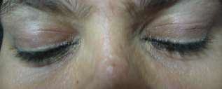
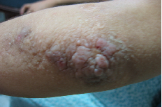
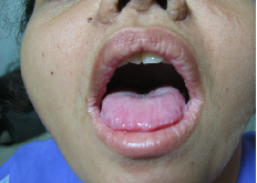
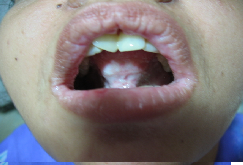
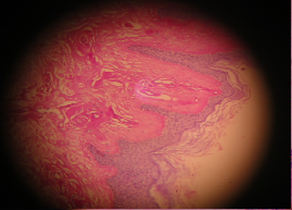
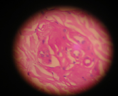
Two sisters ages 16 and 11, born of a second-degree consanguineous marriage, presented with hoarseness at birth, multiple depressed scars on the face, and beaded papules on the margins of the eyelids since early childhood (figs. 1 and 6). Skin-colored, verrucous papules and nodules were present on the elbows (fig. 2), axillae (fig. 7) and sites of friction and movement including hands (fig. 8). The tongue was enlarged and woody, hard on palpation, and the patients were unable to protrude it beyond the margins of the lips (fig 3 and 4). The older girl had diffuse hair loss over the scalp (fig. 5). Dentition was normal. Laryngoscopy revealed hypertrophic tonsils and hypertrophic vocal cords with normal and equal movements. The upper and lower eyelid margins showed the typical papules arranged like beads on a string. Further systemic examination and skull X-rays were normal. The histopathology of a verrucous papule revealed deposition of a pale, homogenous, eosinophilic, hyalinelike material, which was Congo-red-negative and PAS-positive. (figs. 9-12) The blood vessels stood out prominently with hole formation. The material was deposited around blood vessels resulting in an onion-skin appearance and around the eccrine ducts and pilosebaceous units as well.
|
|
| Figure 1 | Figure 2 |
|---|---|
| Skin lesions in 16-year-old girl: Eyelid papules and pocklike scarring (fig. 1). Verrucous papules and nodules over elbow (fig. 2). | |
|
|
| Figure 3 | Figure 4 |
|---|---|
| Skin lesions in 16-year-old girl: Inability to protrude tongue (fig. 3). Infiltration of frenulum and oral mucosae (fig. 4), | |
|
|
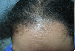
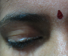
| Figure 5 | Figure 6 |
|---|---|
| Skin lesions in 16-year-old girl: Scarring Alopecia (fig 5). Skin lesions in an 11-year-old girl: Eyelid papules and pocklike scarring (fig. 6). | |
|
|
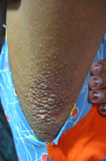
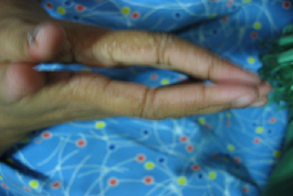
| Figure 7 | Figure 8 |
|---|---|
| Skin lesions in an 11 year-old-girl: Verrucous papules in the axilla (fig. 7). Verrucous papules on the fingers (fig. 8). | |
|
|
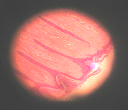
| Figure 9 | Figure 10 |
|---|---|
| Eosinophilic hyaline material consisting of thick, homogenous bundles that extend perpendicular to the skin surface. | |
|
|
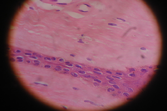
| Figure 11 | Figure 12 |
|---|---|
| PAS-positive material deposited around blood vessels, eccrine ducts, and pilosebaceous unit. | |
Discussion
Hoarseness in infancy, beaded papules along eyelid margins, and woody tongue clinched the diagnosis of lipoid proteinosis. Scars on the face may pose difficulty in differentiating from erythropoietic protoporphyria. Typical histology, absence of photosensitivity, and the presence of skin lesions on covered areas help to delineate this condition. In adults, the differential diagnosis includes lichen myxedema and myxedema. The prognosis of lipoid proteinosis is generally good despite the progressive nature of the disease until early adulthood. Widespread visceral involvement is described, and associated diabetes is reported. Treatment is limited and consists of dissection of vocal cords and dermabrasion for skin lesions. Treatment with Dimethylsulfoxide, D-pencillamine and etretinate has had variable results.
References
1. Black MM. Lipoid proteinosis, In : Textbook of Dermatology, edited by Champion RH, Burton JL, Ebling FJG, 5th edn. Blackwell Scientific Publication, Oxford, 1992, 2347 - 2348.2. Singh G, Misra D. Lipoid proteinosis. Int J Dermatol. 1988 Jun;27(5):344-5. PubMed
3. Yesudian P, Bhasker G. A study of five cases of lipoid proteinosis. Australas J Dermatol. 1972 Aug;13(2):60-8. PubMed
4. Wong CK, Lin Cs. Remarkable response of Lipoid Proteinosis to Oral Dimethylsulfoxide. Br J Dermatol. 1988 Oct;119(4):541-4. PubMed
5. Gruber F, Manestar D, Stasic A, Grgurevic Z. Treatment of lipoid proteinosis with etretinate. Acta Derm Venereol. 1996 Mar;76(2):154-5. No abstract available. PubMed
6. Hamada T. Lipoid Proteinosis Clin Exp Dermatol. 2002 Nov;27(8):624-9. PubMed
7. Kaya TI, Kokturk A, Tursen U, Ikizoglu G, Polat A. D-penicillamine treatment for lipoid proteinosis. Pediatr Dermatol. 2002 Jul-Aug;19(4):359-62. PubMed
8. Touart DM, Sau P. Cutaneous deposition diseases. Part I. J Am Acad Dermatol. 1998 Aug;39(2 Pt 1):149-71; PubMed
© 2003 Dermatology Online Journal