Lupus vulgaris diagnosed after 37 years: A case of delayed diagnosis
Published Web Location
https://doi.org/10.5070/D39k34f7gxMain Content
Lupus vulgaris diagnosed after 37 years: A case of delayed diagnosis
Enver Turan1 MD, Nurdan Yurt2 MD, Yavuz Yesilova1 MD, Ozgur Ilhan Celik3 MD
Dermatology Online Journal 18 (5): 13
1. Department of Dermatology, Faculty of Medicine, University of Harran, Sanliurfa, Turkey2. Dermatology Clinic, Gumushane Government Hospital, Gumushane, Turkey
3. Department of Pathology, Ministry of Health Batman Regional Government Hospital, Batman, Turkey
Abstract
Lupus vulgaris is the most common chronic, progressive form of cutaneous tuberculosis. Lesions are generally solitary and found on the head and neck region. Cutaneous tuberculosis can present with different clinical appearances. Therefore, it does not necessarily have characteristic findings and can be difficult to diagnose. Although there were typical clinical findings, the diagnosis of our case was delayed because of its asymptomatic course.
 |  |
| Figure 1 | Figure 2 |
|---|---|
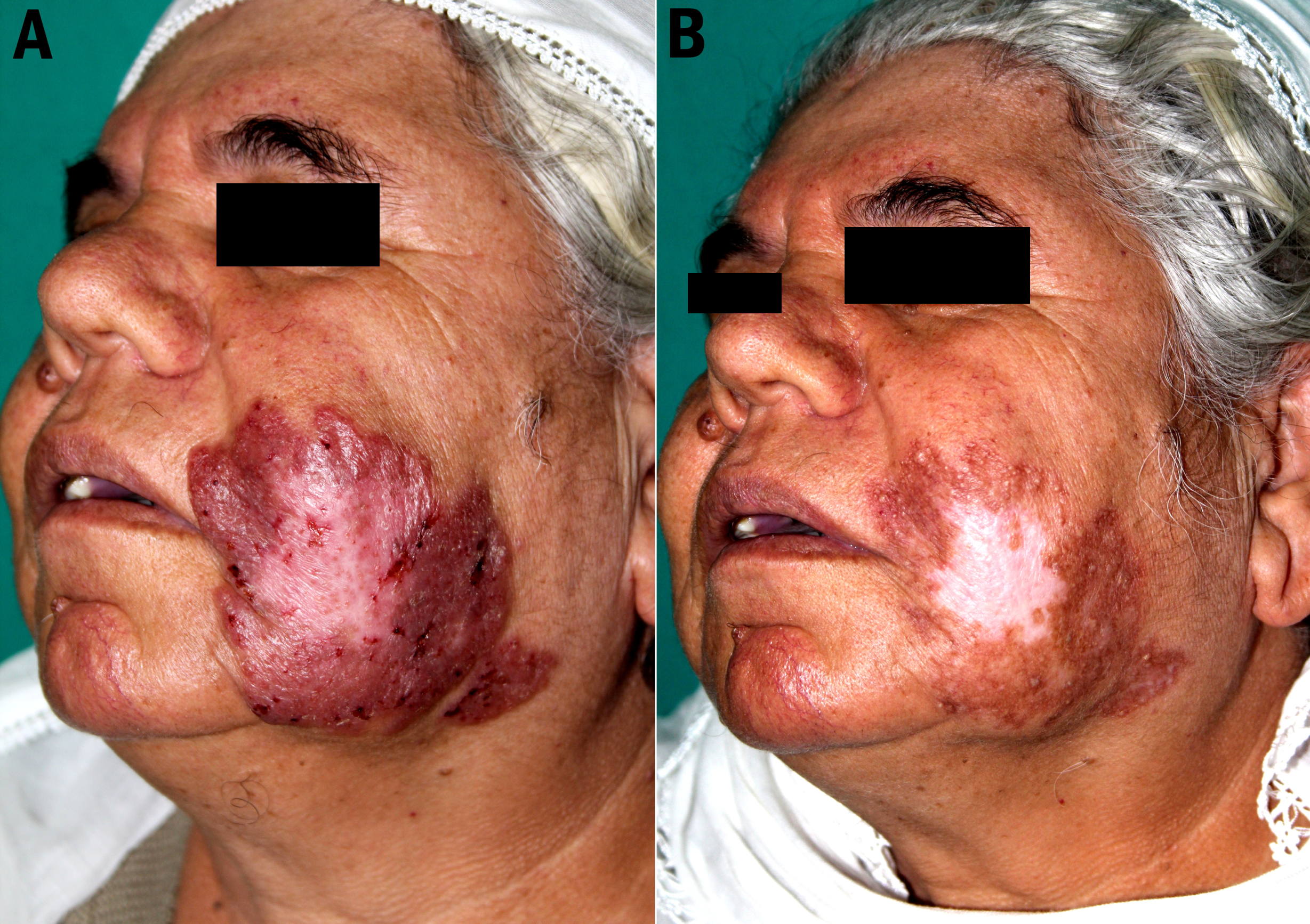
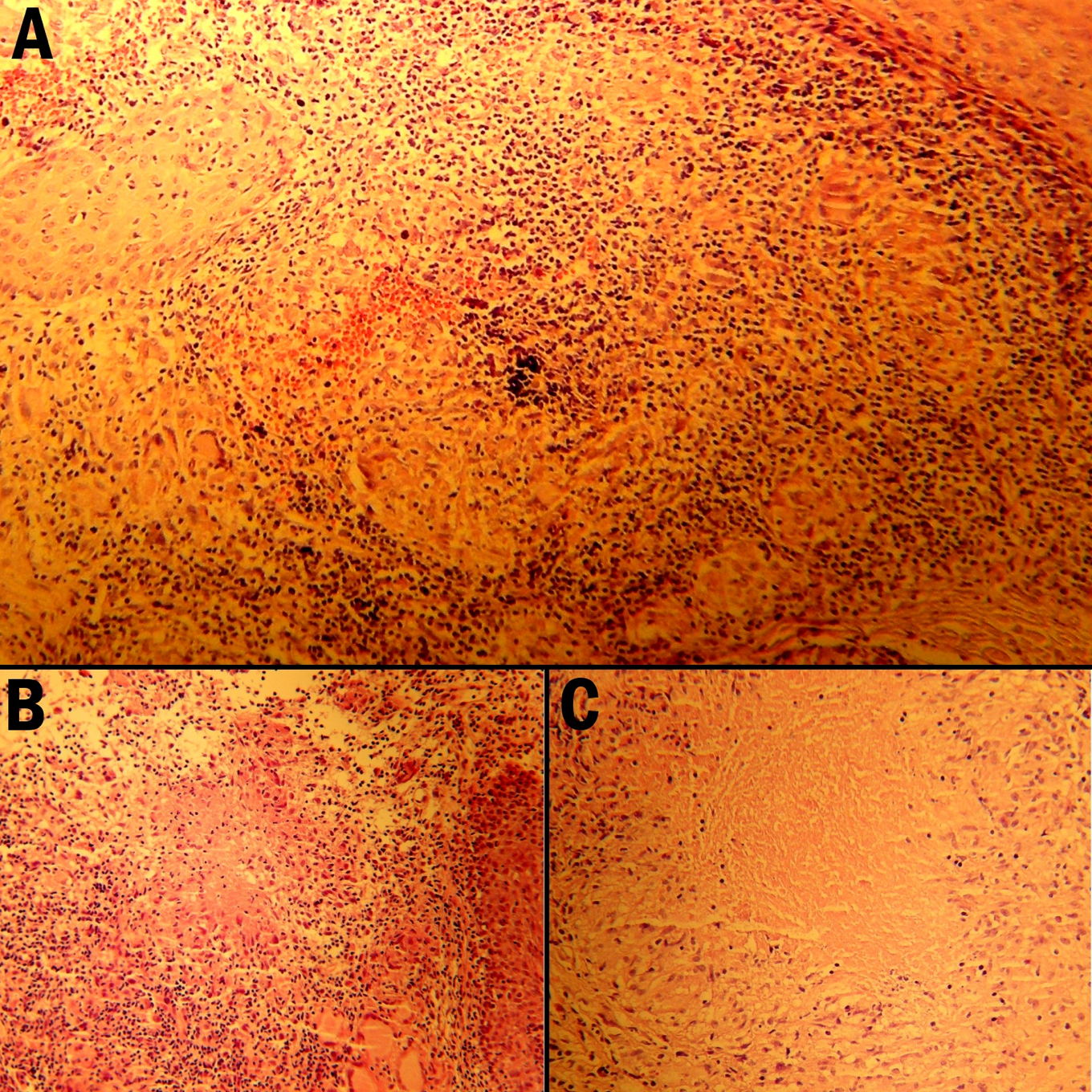
| Figure 1A. Well-demarcated, large, reddish-brown plaque with an atrophic center located on the left cheek. Figure 1B. The lesion healed with hypopigmented, atrophic scarring at the end of the 6 months of treatment. Figure 2A and Figure 2B. Granulomatous inflammation in the dermis, which had Langhans type giant cells and epithelioid histiocytes in the center and was surrounded by plasma cells and lymphocytes (H&E, x100). Figure 2C. Langhans type giant cells, epithelioid histiocytes, and caseation necrosis within the granulomas (H&E, x400). | |
A 59-year-old woman presented to our clinic with an erythematous, scaly plaque on her left cheek existing for 37 years. Clinical examination demonstrated a large, well-defined, reddish-brown plaque with an atrophic center, located on the left cheek (Figure 1A). Morphology of the lesion and apple jelly color on diascopy suggested lupus vulgaris. There was no past history of tuberculosis and no known exposure to tuberculosis. She had no previous BCG vaccination. Otherwise, she was healthy and had no complaints.
Hematological and radiological investigations were normal. Polymerase chain reaction from the tissue specimen for M. tuberculosis was positive and a Mantoux test was measured to be 22 mm. Tissue culture and Ziehl-Neelsen stain for tubercle bacilli and Grocott stain for fungus were negative. Histopathological examination of the specimen taken from the lesion revealed a granulomatous infiltration in the dermis, which had Langhans type giant cells and epitheloid cells in the center and was surrounded by plasma cells and lymphocytes. There were foci of caseation necrosis within the granulomas (Figure 2). The patient was diagnosed with lupus vulgaris and treated with isoniazid, rifampicin, ethambutol, and pyrazinamide therapy for 4 months followed by isoniazid and rifampicin for 2 months. The lesion healed with hypopigmented, atrophic scarring at the end of the 6 months (Figure 1B).
Lupus vulgaris is the most common chronic, progressive form of cutaneous tuberculosis. Lesions are generally solitary and found on the head and neck region. Cutaneous tuberculosis can present with various clinical appearances. Therefore, findings may not be characteristic and difficult to diagnose. Although there were typical clinical findings, the diagnosis of our case is delayed because of the asymptomatic course [1].
Although demonstration of mycobacteria is necessary for definitive diagnosis of cutaneous tuberculosis, generally it is not possible because it is paucibacillary. Mycobacterium tuberculosis often fails to grow in culture, especially when taken from chronic lesions or patients having a high degree of immunity. For growth of bacilli on routine culture media like Lowenstein-Jensen or Middlebrook, it takes nearly 3-4 weeks but this period decreases to 10-14 days for the BACTEC radiometric system. Polymerase chain reaction is a fast, sensitive, and specific diagnostic method for paucibacillary disease like lupus vulgaris [2, 3]. In our case, DNA of Mycobacterium tuberculosis was positive with PCR technique. However, no growth on the culture medium was observed.
Combination therapy should be done in cutaneous tuberculoses like lupus vulgaris, tuberculosis verrucosa cutis, and scrofuloderma. Multidrug therapy is required for the treatment of cutaneous tuberculosis so that resistance and recurrences can be prevented [1, 4].
In conclusion, tuberculosis should be suspected in cases of chronic skin lesions which are resistant to conventional antibiotic therapy and show no growth on culture.
References
1. Barbagallo J, Tager P, Ingleton R, Hirsch RJ, Weinberg JM: Cutaneous tuberculosis: diagnosis and treatment. Am J Clin Dermatol 2002, 3(5):319-328. [PubMed]2. Negi SS, Basir SF, Gupta S, Pasha ST, Khare S, Lal S: Comparative study of PCR, smear examination and culture for diagnosis of cutaneous tuberculosis. J Commun Dis 2005, 37(2):83-92. [PubMed]
3. Almaguer-Chavez J, Ocampo-Candiani J, Rendon A: Current panorama in the diagnosis of cutaneous tuberculosis. [Spanish] Actas Dermosifiliogr 2009, 100(7):562-570. [PubMed]
4. Rama Rao GR, Narayan BL, Amareswar A, Sandhya S: Directly observed treatment short course and cutaneous tuberculosis: our experience. Indian J Dermatol Venereol Leprol 2011, 77(3):330-332. [PubMed]
© 2012 Dermatology Online Journal